Quantcast
Channel: Endocrinology – Geeky Medics
Viewing all 51 articles
Browse latest View live

Type 1 Diabetes Mellitus

$
0
0
What is Type 1 Diabetes Mellitus? 

Type 1 Diabetes Mellitus is a form of diabetes that occurs due to autoimmune destruction of the insulin producing pancreatic beta cells. The loss of insulin producing cells leads to insulin deficiency which in turn causes hyperglycaemia. Patients with this disease will always require insulin to control blood glucose levels. Without treatment patients develop diabetic ketoacidosis which can progress to coma and death. It is therefore essential to recognise signs of type 1 diabetes as early as possible to allow prompt treatment.


The disease commonly affects young otherwise healthy individuals however it can occur at any age. The disease has been associated with other autoimmune diseases such as thyroid disease, coeliac’s disease, pernicious anaemia, vitiligo and others. It is thought to be the result of a genetic susceptibility combined with exposure to an environmental trigger (e.g. virus)

Causes
Genetic

Over 90% carry HLA DR3 +/- DR4 suggesting these genes add significant risk of disease

The gene IDDM1 is thought to be strongly associated with development of type 1 diabetes

Environmental

Studies of identical twins have shown only 30% concordance of developing type 1 diabetes.
This suggests that the environment plays a very important role in the development of the disease
It has been suggested that a virus may trigger the disease in genetically suceptible individuals
This is thought to occur due to a virus having similar antigens to the pancreatic beta cells causing inappropriate immune activation. The Coxsackie virus is one organism which has been implicated as a trigger in some cases

Symptoms & Signs
Symptoms

Polyuria -frequent urination
Polydipsia -increased thirst

Polyphagiaincreased hunger
Weight loss -
unrelated to diet
Lethargy
Genital thrush
Dehydration

Blurred Vision
Nausea
Abdominal Pain

Signs

Hyperglycaemia
Glucosuria - glucose in urine
Ketone bodies
Diabetic retinopathy
Peripheral Neuropathy
Foot ulcers
Pear drop scented breath -ketoacidosis

Diabetic Ketoacidosis

Diabetic Ketoacidosis (DKA) is a potentially life threatening condition most often seen in type 1 diabetics. It occurs due to a lack of insulin, which prevents the bodies cells from absorbing and utilising any glucose in the blood. As a result the body switches it’s metabolism to breakdown fat to produce energy. This process produces acidic ketones as a byproduct and these build up in the blood causing a metabolic acidosis. Left untreated the condition ultimately leads to coma and death, therefore signs need to be recognised early and treated quickly.

Clinical features

Nausea/Vomiting
Severe abdominal pain
Dehydration
Breath smells of pear drops
High Ketones
Severe Hyperglycaemia
Severe Hyperkalaemia
Kussmaul Respiration - deep gasping breathing
Cerebral Oedema
Coma

Treatment

The main aims of treatment are to reduce blood glucose & ketone levels using insulin whilst correcting dehydration & other electrolyte abnormalities:

  • Insulin
  • Potassium
  • Bicarbonate
  • IV Saline
Investigations

Diagnosis is made based upon:

  • Classical type 1 diabetes symptomsweight loss, ketoacidosis, polyuria, polydipsea etc
  • Hyperglycaemia - (fasting glucose >7 mmol/L)  or (random glucose >11.1 mmol/L)

.

Ketones - can be checked to assess if patient has ketoacidosis

Antibodies – shouldn’t be used for diagnosis but may be helpful in differentiating type 1 & type 2

Management

All patients with type 1 diabetes require insulin replacement therapy

Patients must regularly check their blood glucose levels & adjust their doses accordingly

 
Subcutaneous Insulins 

Very Fast Acting – usually injected at start of meal to match what is eaten – e.g.Novorapid


Combination Insulin - Fast & long acting insulin mixed together - e.g. Novomix 30% fast 70% long


Long acting - acts over long period of time therefore good for night time - e.g. Glargine


Regimes

The regime of treatment is influenced by the type of lifestyle an individual has:

  • Biphasic regime – twice daily combination insulin -useful if patient has a predictable lifestyle
  • QDS regime – use Novorapid before meals & have 1 dose of long acting insulin at night – flexible
 
Insulin Pump Therapy

Insulin pump therapy aims to more closely mimic the function of a healthy pancreas

The pump uses fast acting insulin such as Novorapid

It involves having a small cannula placed in the subcutaneous fat, usually around the waist

This is connected to the insulin pump which provides the insulin infusion & boluses

.

How it works

The pump releases a constant rate of insulin throughout a 24hr day, known as the basal rate

This can be altered in real time by the patient depending on their needs50% increase when ill

.

The pump can also give a bolus of insulin before each meal, this involves:

  • Patient calculating the amount of carbohydrates in a meal
  • Then calculating amount of insulin required depending on their insulin:carb ratio
  • Setting the pump to provide the required units of insulin over a short time period e.g. 15 mins
 

Insulin pumps give individuals much more precise control over their blood glucose level.

This is empowering for the patient & over time they learn which settings work best for them

Insulin pumps have been shown to improve quality of life & lower HBA1C if used appropriately

There is an increased risk of DKA due to using  only rapidly acting insulin

.

NICE recommends insulin pump therapy for type 1 diabetes patients who:

  • Suffer repeated & unpredictable episodes of severe hypoglycaemia when trying to reach acceptable HBA1C
  • HBA1C levels have remained above 8.5% with multiple daily injections
  • In children less than 12 years where multiple daily injections is impractical or innappropriate
Eye Screening

There is now an annual national eye screening program for all people with diabetes
This aims to recognise and treat diabetic retinopathy at an early stage before vision is lost

Podiatry

Its essential for those with diabetes to go to regular appointments with a Podiatrist
Diabetes can cause peripheral neuropathy & vascular insufficiency which can lead to ulcers
The aim is to detect early signs of foot disease and take measures to prevent progression
This involves advice on foot care and appropriate foot wear among other things

HBA1C monitoring

The patient will have their HBA1C monitored by their doctor
The HBA1C shows the level of blood glucose control over the previous 3 months
It can identify patients with poor control and whose treatments may have become ineffective
It is normally recommended that a patient keep their HBA1C below 7.5%

Prognosis

The longer an individual has diabetes the more risk they have of developing complications:

  • Peripheral Neuropathy
  • Diabetic Retinopathy
  • Chronic Renal Failure
  • Cardiovascular disease - MI
 

The risk of developing these complications can be largely reduced by good blood glucose control.  Therefore the probability of each of these depends on the individual patient & co-morbidities


Addison’s Disease

$
0
0
What is Addison’s disease?

Addison’s disease involves the underproduction of cortisol & aldosterone by the adrenal cortex

The disease is very rare with around 8 in a million people developing the disease.

It generally affects individuals between the ages of 20-50 however it can occur at any age.

It’s essential you first understand the hypothalamic-pituitary-adrenal axis in normal individuals

It will then be easier to understand how this system goes wrong in disease

So check out the adrenal axis article first which can be found here

What does the adrenal cortex do?

The Adrenal Cortex is responsible for producing Cortisol and Aldosterone 

Cortisol 

Cortisol is a steroid hormone (glucocorticoid)which is released under stress & low steroid levels

It’s main function is to increase blood glucose levels by promoting gluconeogenesis

Cortisol also suppresses the immune system & increases fat, protein & carb metabolism


Random Fact!

You may recognise cortisol’s pharmaceutical name which is Hydrocortisone
Hydrocortisone is used to reduce inflammation leveraging Cortisol’s immunosuppresive effect
.
 
Aldosterone 

Aldosterone is also a steroid hormone (mineralocorticoid )

It’s main function is to increase blood volume

It causes reabsorption of sodium and water as well as causing excretion of potassium


Random Fact!

Drugs that interfere with the secretion or action of Aldosterone are in use as antihypertensives
An example is Spironolactone which blocks Aldosterone receptors
Causes
Autoimmune destruction

Autoimmune destruction of the adrenal cortex is the most common cause of Addison’s disease- 70%

Antibodies are produced against the adrenal cortex or an enzyme called 21-hydroxylase

These antibodies cause destruction of the adrenal cortex & deficiency in cortisol & aldosterone


A rarer disorder known as Autoimmune Polyglandular Deficiency can also cause addison’s disease

This disease occurs due to autoimmune attack against multiple endocrine organs

These includeparathyroid, adrenal, thyroid, pituitary, pancreas

These disorders are genetic in origin and often involve the Auto Immune Regulator Gene (AIRE)


Genetic Disorders

Adrenoleukodystrophy (Schilder’s disease)
A rare, inherited disorder of fatty acid metabolism
Presents in childhood
Leads to progressive brain damage, failure of the adrenal glands & eventually death

Malignancy

Adrenal Metastasis - common in lung, breast & kidney cancer

However very few result in development of addison’s disease


Infection 

Tuberculosis -  in rare circumstances it can spread to adrenals and destroy the cortex

Opportunistic infections – cryptococcosis, candidiasis, histoplasmosis (common in AIDS)

 
Vascular

Waterhouse-Friderichsen syndrome:

  • Massive haemorrhage of adrenals
  • Usually due to meningococcal septicaemia.

Infarction – a thrombus from another site occludes blood supply of adrenal gland

 
Adrenal dysgenesis

Very rare

Almost always caused by genetic mutations

Congenital Adrenal Hypoplasia is an example – caused by mutations in DAX1 gene

 
Impaired Steroid Production

The adrenal glands produce steroid hormones from cholesterol you consume

This process involves many enzymes, and a mutation in any of these can cause problems

Congenital Adrenal Hyperplasia is caused by mutations in some of these enzymes

 
Infiltration

Haemochromotosis:

- Disorder of increased intestinal iron absorption
- Leading to high levels of body iron
- This excess iron is deposited in the adrenal tissue as well as the liver, heart, pancreas etc
- The iron accumulates and becomes toxic causing death of the adrenal tissue


Iatrogenic (caused by doctors!)

Adrenal haemorrhage as a result of anticoagulant therapy

Removal of adrenal glandse.g. in malignancy

Fluconazole & Ketoconazole inhibit cortisol production

Phenytoin & Rifampicin induce enzymes which increase cortisol metabolism by the liver

Signs & Symptoms

Anorexia & Weight Loss – 90% of cases

Fatigue & Lethargy

.

Hyperpigmentation;

- Due to increased POMC
- Even in areas not exposed to sun
- Hand creases, Nipple, Buccal Mucosa (pathognomonic)
- Darkening of scars

.

Generalised Weakness

GI symptoms - nausea, vomiting, diarrhoea

Postural Hypotension - due to lack of aldosterone

Decreased libido in males

Calcification of the Pinna of the Ear - in longstanding disease – rare but amazing if you spot it!

Decreased axillary & pubic hair

Symptoms of other endocrine disease - due to possibility of autoimmune polyendocrine syndrome

Addisonian Crisis

Addisonian crisis is a collection of serious symptoms which indicate severe adrenal insufficiency

This is a medical emergency and if not managed promptly it can lead to death

Causes

An individual with undiagnosed addison’s disease,

A patient not adhering to treatment

Any other problem that has caused sudden loss of adrenal function (haemorrhage, infection)

 
Symptoms

Hypotension

Severe abdominal pain

Severe Vomiting & Diarrhoea

High fever

Shock

Confusion

Convulsions

Death

 
Management 

Clinical suspicion + a low serum cortisol is sufficient to diagnose

Do not delay treating by waiting for confirmatory tests

Treat dehydration with 0.9% saline (beware of worsening electrolyte disturbances)

High doses of hydrocortisone should be given

Glucose may be required if hypoglycaemic

Investigate the cause of the crisis and attempt to treat it

Investigations
Biochemical tests

Common abnormalities include:

  • Hypercalcaemia
  • Hypoglycaemia
  • Hyponatraemia
  • Hyperkalaemia
  • Eosinophilia
  • Metabolic Acidosis
 
Autoantibodies

Adrenal Cortex Antibodies

21-Hydroxylase antibodiespresent in 80%

 
Imaging

CT abdomen

Atrophic adrenal glands seen in autoimmune adrenalitis

Enlarged adrenals seen in infective, infiltrative and metastatic causes

Diagnosis
ACTH Stimulation Test 

This involves giving the patient an IM injection of synthetic ACTH (Synacthen)

Cortisol is measured at 30 & 60 mins then at 2, 4, 8, 12, 24 hours

Serum cortisol should rise substantially during this period of time

In Addison’s disease cortisol will not rise

In secondary adrenal failure the cortisol will steadily begin to rise

Management

The treatment strategy is to replace the hormones the adrenal cortex produces

Cortisol (glucocorticoid) replacement

Hydrocortisone is the treatment of choice for cortisol replacement

It is reliable due to it’s predictable absorption rate

It is given in 3 daily doses (morning, midday and evening) to mimic the bodies circadian rhythm

 
Aldosterone (mineralocorticoid) replacement

Fludrocortisone is the treatment of choice for aldosterone replacement

The dose is usually around 100mcg once daily

 
DHEA replacement

DHEA is also produced by the adrenal cortex

It is a precursor of oestrogen and testosterone

It also acts as a weak androgen itself

Replacement can improve mood and general well being

Prognosis
Individual who receive adequate therapy should live a normal healthy life

However if not managed properly addisonian crisis can lead to death

How the Adrenal Axis Works

$
0
0
What is the Adrenal Axis?

The adrenal axis (hypothalamic-pituitary-adrenal axis) refers to a complex set of interactions and feedback loops between the hypothalamus, pituitary and adrenal glands. This system regulates the bodies response to stress, immune function, energy expenditure, mood, emotions & libido. It is therefore an incredibly important body system which can cause huge problems if it malfunctions

What are the adrenal glands?

There is an Adrenal gland located above each kidney

They both produce a number of hormones

The Adrenal glands are composed of a outer “Cortex” and an inner “Medulla”

Adrenal Cortex

The Adrenal Cortex is responsible for producing Cortisol and Aldosterone

Cortisol 

Cortisol is a steroid hormone (glucocorticoid)which is released under stress & low steroid levels

It’s main function is to increase blood glucose levels by promoting gluconeogenesis

Cortisol also suppresses the immune system & increases fat, protein & carb metabolism

Random Fact!

You may recognise cortisol’s pharmaceutical name which is Hydrocortisone
Hydrocortisone is used to reduce inflammation leveraging Cortisol’s immunosuppresive effect

.
Aldosterone

Aldosterone is also a steroid hormone (mineralocorticoid)

It’s main function is to increase blood volume

It causes reabsorption of sodium and water as well as causing excretion of potassium


Random Fact!

Drugs that interfere with the secretion or action of Aldosterone are in used as antihypertensives

An example is Spironolactone which blocks Aldosterone receptors
Adrenal Medulla

The adrenal medulla produces adrenalinenoradreniline (both have similar actions)

They are released as an acute response to stress & imitate many of the effects of cortisol

They essentially prepare the body for “fight or flight”↑HR,  ↑BP,  ↑Blood flow to muscles

Their release is under direct control from the hypothalamus – allowing  fast response to stress

Adrenaline & Noradreniline’s effects are not as long lasting as cortisol

How Cortisol is Regulated
  1. Corticotrophic releasing hormone (CRH) is secreted from the Hypothalamus
  2. This release is influenced by stress levels, time of day & serum cortisol levels
  3. CRH travels in the blood & binds to specific receptors on the Pituitary Gland
  4. This binding causes increased production of ACTH (adrenocorticotrophic hormone)
  5. ACTH is released into the blood stream where it travels to the Adrenal glands
  6. ACTH binds to specific receptors on the Adrenal Cortex
  7. This stimulates the Adrenal Cortex to release Cortisol into the blood
  8. Cortisol enables the body to cope with stress in a more effective manner
  9. Increased levels of Cortisol also have an immunosuppresive effect
  10. Blood glucose is also increased via breakdown of glycogen, protein & fat
  11. Increased serum Cortisol inhibits production of CRH & ACTH via negative feedback


.
.
How Aldosterone is Regulated

Low blood volume triggers release of Angiotensin II
Angiotensin II stimulates the Adrenal Cortex to produce Aldosterone
Aldosterone has a number of different actions;

  • Sodium Reabsorption
  • Water Reabsorption
  • Potassium Excretion

Aldosterone’s actions result in a net increase in blood volume
Increased blood volume inhibits production of Angiotensin II via negative feedback


Cushing’s Syndrome

$
0
0
What is Cushing’s Syndrome? 

Cushing’s Syndrome refers to a disease process caused by abnormally high levels of Cortisol

This can result from overdose of glucocorticoid drugs or from tumours producing Cortisol or ACTH

Cushing’s Syndrome has a high mortality if left untreated – 50% at 5 years

It most commonly affects individual between the ages of 20-40 years

It is a rare disorder with 2 in a million people developing it

Women have a significantly higher risk of developing Cushing’s Syndrome than men do

Make sure to read the “How the Adrenal Axis Works” article before reading about Cushing’s Syndrome

Causes
The causes of Cushing’s syndrome can be divided into;

Exogenous (caused by hormones outside the body e.g. iatrogenic)

Endogenous (caused by hormones produced by the body itself e.g. pituitary, adrenal, ectopic)
.
Iatrogenic

The most common cause of Cushing’s Syndrome is glucocorticoid treatment

Steroids are used in many diseases to reduce inflammation & suppress immune function

Diseases requiring steroids include asthma, autoimmune disease & organ transplantation

Long term use of steroids cause the symptoms of cushing’s syndrome

.
 
Pituitary Cushing’s (Cushing’s Disease)

Pituitary Cushing’s results from a benign pituitary adenoma secreting excess levels of ACTH

High levels of ACTH in turn caused increased release of Cortisol from the Adrenal Cortex

Cushing’s Disease is the commonest endogenous cause of Cushing’s Syndrome – 70%

.
 
Adrenal Cushing’s

Adrenal Cushing’s involves over production of Cortisol directly by the Adrenal Cortex

This can be due to Adrenal Adenoma’s or Hyperplasia of the Adrenal Glands

The Adenoma or increased Adrenal Tissue release excessive amounts of Cortisol

These high levels of Cortisol cause the characteristic symptoms of Cushing’s Syndrome

.
 
Ectopic Cushing’s
 

Ectopic Cushing’s is due to ↑ production of ACTH from a tumour outside of the adrenal axis

Most commonly this type of Cushing’s is seen in Small Cell Lung Cancer

The tumour for some unknown reason has genes switched on which enable it to produce ACTH

This stimulates the Adrenal Cortex to produce increased levels of Cortisol

High levels of Cortisol give rise to the symptoms of Cushing’s Syndrome


Pseudo Cushing’s Syndrome

Pseudo Cushing’s refers to a condition in which a patient has the signs & symptoms of cushing’s

However the cause is not related to the hypothalamic-pituitary-adrenal axis

Pseudo Cushing’s Syndrome’s cause is therefore idiopathic

Patients often have a raised total serum Cortisol & ACTH

The Cortisol levels also usually fail to be suppressed by Dexamethasome

.

Common Causes of Pseudo Cushing’s Syndrome include;

  • Alcoholism
  • Depression
  • Stress
  • Eating Disorders

Often Pseudo-Cushing’s Syndrome spontaneously resolves

Particularly when Alcoholics undergo abstinence

Signs & Symptoms

Central Obesity- sparing of limbs

Moon face – rounded facial appearance

Buffalo Hump – fat pad on back of neck

Acne Thinning of skin - cracks, splits & bruises easily

Dry skin

Telangiectasia

Excessive sweating

Stretch marks red striae around abdomen, hips

Proximal muscle weakness

Osteoporosis

Hirutism

Amenorrhea

Insomnia

Euphoria – common side effect of high dose steroids

Psychosis – uncommon

Diagnosis
Dexamethasome Suppression test

- Give 1mg of oral dexamethasome at midnight
- Record serum cortisol before & at 8am
- Cortisol & ACTH should decrease due to negative feedback in normal individuals
- In Cushing’s Cortisol will not be decreased

.

Further testing if suppression test positive

- CT of adrenal glandsadrenal adenoma, malignancy
- MRI of pituitary fossapituitary adenoma/ malignancy
- Search for ectopic ACTH production - CT Neck, Thorax, Abdomen

Management
Iatrogenic Cushing’s Syndrome

In Iatrogenic Cushing’s slowly reduce the dose of the glucocorticoid

Aim to stop glucocorticoid treatment if at all possible

Cushing’s syndrome should then resolve

.
Pituitary/ Adrenal Adenoma

Pituitary adenomas can be removed via trans-sphenoidal surgery

Adrenal Adenoma’s can be removed via Adrenalectomy

It is essential to replace corticosteroids at least short term

ACTH takes time to recover normal levels of production after long term suppression

Failure to give replacement therapy can result in Adrenal Crisis

.
Unknown cause

Sometimes the cause for high cortisol levels cannot be determined

In these cases it may be necessary to perform a bilateral adrenalectomy

This is a last resort with the significant risk of developing Nelsons Syndrome

Nelson Syndrome involves:

  • Rapid growth of a pre-existing pituitary adenoma
  • Due to lack of negative feedback from Cortisol
  • Pituitary irradiation is sometimes performed to prevent this
.

Lifelong Hydrocortisone therapy will be required after bilateral adrenalectomy

Prognosis

Left untreated cushing’s syndrome has a high mortality – 50% at 5 years

The prognosis is good for those who recieve adequate treatment

However issues may persist such as diabetes, hypertension & osteoporosis

Individuals would usually have regular review by an endocrinologist

.

Type 2 Diabetes Mellitus

$
0
0
What is Type 2 Diabetes Mellitus?

Type 2 Diabetes Mellitus is a disorder of metabolism characterised by high levels of glucose in the blood as a result of insulin resistance and insulin deficiency.  The disease is much more common in those who are overweight or obese and it often runs in families suggesting a significant genetic component. It tends to occur in people older than 40, however increasingly younger individuals are developing the disease.  In the UK around 1 in 20 over 65′s and around 1 in 5 over 85′s have diabetes.  In developed nations the prevalence of diabetes is increasing rapidly, due to changing environmental and lifestyle factors.

Causes

Diabetes is caused by a combination of environmental & genetic factors

Factors increasing risk of type 2 diabetes

Overweight/Obesity

Age – increasing risk with age

Poor diet

Lack of  exercise

Large waist – >31.5 inches (woman), >37 inches (man)

First degree relative with type 2 diabetes – adds significant risk

South Asian Origin – 6x more risk than white caucasian

Impaired Glucose Tolerance

Other medical conditions - acromegaly, cushing’s, pancreatitis, metabolic syndrome

Medicationse.g. long term steroids

Signs & Symptoms
Symptoms

Polyuria

Polydipsia

Unexplained weight loss

Visual Blurring

Lethargy

Genital Thrushcandida

 
Signs

Peripheral neuropathy - glove & stocking

Poor peripheral pulses

Foot Ulcersdue to poor blood supply + neuropathy

Diabetic Retinopathyoedema & bleeding in back of the eye

Proteinurea – diabetic nephropathy

Acanthosis Nigricans – darkening of skin folds around neck/axilla (insulin resistance)

Necrobiosis Lipodica - shiny reddish brown pigmentation on legs

Diagnosis

Symptoms of type 2 diabetes as well as one of the following:

  • Fasting glucose ≥ 7mmol/L
  • Random glucose ≥ 11.1mmol/L
  • Oral Glucose Tolerance Test ≥11.1mmol/L

If no symptoms present raised glucose should be demonstrated on 2 occasions

Lifestyle Changes

In it’s early stages type 2 diabetes can often be treated by altering lifestyle factors

Examples of this include:

- Weight Loss Promoting, Low Fat, Low GI diet
- Doing regular aerobic exercise
- Doing resistance training with weights

Patients who follow these steps are more likely to avoid needing drug therapy

Drug Therapy
Metformin (biguanides)

Metformin is the 1st line therapy for overweight/obese patients with type 2 diabetes

It increases insulin sensitivity, allowing their endogenous insulin to better do it’s job

The drug decreases hepatic gluconeogenesis whilst increasing muscle glucose uptake


In the long term a reduction in HBA1C is usually seen in most patients

It has been shown to significantly reduce mortality in overweight patients

Common side effects are GI upset with flatulencediarrhoea, cramps

These tend to subside after a few weeks treatment


Sulfonylureas

Sulfonylureas are used as 1st line in non-obese patients with type 2 diabetes

Gliclazide is one of the most commonly used of this drug class

It works by binding to pancreatic beta cells & causing increased release of insulin

This drug typically results in a 1-2% reduction in HBA1C in the long term

Side effects include hypoglycaemia due to excess insulin release & also weight gain

.
Thiazolidinediones

Thiazolidinediones causes insulin sensitisation

Rosiglitazone is one of the most commonly used agents of this class

They are usually given in combination with metformin or a sulphonylurea


The drug binds to PPAR’s (peroxisome proliferator-activated receptors)

When PPAR’s are activated they cause transcription of a number of genes

These activated genes ultimately result in decreased insulin resistance


Thiazolidinediones cause around a 0.5-1.5% reduction in HBA1C

Side effects include fluid retentionweight gain & hepatotoxicity

.
GLP1 Mimetic’s

GLP1 mimetics imitate the effects of GLP1

GLP1 is a hormone produce by intestinal cells when exposed to carbohydrate

It causes insulin secretion, inhibition of glucagon and decreased insulin resistance

It has a short half life lasting only 2 minutes

It only functions when blood glucose is high, meaning it does not cause hypoglycaemia


Exenatide is a GLP1 mimetic currenltly used

It is administered as a twice daily injection

It results in a 0.8% improvement of HBA1C over 12 months


Insulin

Some patients with type 2 diabetes require insulin to improve glycaemic control

Insulin directly reduces blood glucose by increasing cellular uptake of glucose

A patient will therefore be required to check their blood glucose regularly

The major side effects are hypoglycaemia, weight gain & injection site reactions


Types of Insulin include:

Short acting (Novorapid)

Intermediate acting

Long acting (Glargine)

.

There are many different insulin regimes available

The type of regime is chosen based upon an individuals lifestyle & preferences


Insulin Regimes include:

Basal bolus regime - 3 pre-meal short acting insulin doses + a long acting dose at bedtime

Twice daily - mixture of short & long acting insulin taken at breakfast & tea time

Preventing Complications

In the long term people with type 2 diabetes are at risk of serious complications

These can be reduced and prevented in a number of ways

Monitoring HBA1C

HBA1C is a measure of the proportion of red blood cells that are glycosylated

It allows blood glucose control over the previous 3 months to be assessed

It should be less than 7.5%

This can inform a doctor if a patients treatment is working or if it should be altered

The lower a patients HBA1C the less chance of long term complications


Diabetic foot clinics

Patients with diabetes are at increased risk of developing foot ulcers

This is due to a combination of poor vascular supply & reduced sensation

Therefore it’s important to monitor their feet & this is done in diabetic foot clinics

It involves a podiatrist examining the feet looking for early signs of disease

The podiatrist can then give advice about foot care and arrange any treatment required


Eye screening

Patients with type 2 diabetes are at significant risk of developing eye disease

This is known as diabetic retinopathy and without treatment can result in blindness

There is now a national annual screening program for all diabetic patients

It allows early identification of eye disease and prompt treatment e.g. laser therapy

.

Urine Dipstick

Diabetic nephropathy is a major cause of premature death

1 in 6 patients receiving dialysis will have diabetes

If a dipstick shows a patients urine to be positive for protein it requires further investigation

.

Reducing other complications

Patients are at increased risk of other complications such as cardiovascular disease

These other risk factors are managed by the patients GP e.g. treatment of hypertension

How the Gonadal Axis Works

$
0
0
What is the gonadal axis?

The gonadal axis comprises of the interaction between the hypothalamus, pituitary gland and the gonads.  The system works together to regulate development, reproductionageing and many other body processes.  It’s regulation relies upon a number of complex negative feedback loops which when lost result in disease.

How the male gonadal axis works

1. The hypothalamus secretes GnRH

2. GnRH travels down to the anterior pituitary gland
3. It binds to receptors on the pituitary gland
4. This causes release of LH (luteinizing hormone) & FSH (follicle stimulating hormone)
5. LH & FSH travel in the blood stream to the testicles
6. LH stimulates Leydig cells in the testicles to produce testosterone
  • Testosterone is required for spermatogenesis and many other important biological processes
7. FSH stimulates Sertoli cells to produce androgen binding globulin (ABG) & inhibin
  • ABG is a protein which binds to testosterone & keeps it within the seminiferous tubules
  • Inhibin helps support spermatogenesis and inhibits production of FSH, LH and GnRH

8. levels of testosterone & inhibin cause -ve feedback on the pituitary & hypothalamus


9. This results in decreased production of LH & FSH


10. As a result production of testosterone & inhibin is also decreased


How the female gonadal axis works

1. The hypothalamus secretes GnRH

2. GnRH travels down to the anterior pituitary gland

3. It binds to receptors on the pituitary gland

4. This causes release of LH (luteinizing hormone) & FSH (follicle stimulating hormone)

5. LH & FSH travel in the blood stream to the ovaries

6. When LH & FSH bind to the ovaries they stimulate production of oestrogen & inhibin

  • Oestrogen helps regulate the menstrual cycle & is essential in many body processes
  • Inhibin causes inhibition of activin which is usually responsible for stimulating GnRH production

7. Increasing levels of oestrogen & inhibin cause -ve feedback on the pituitary & hypothalamus

8. This leads to decreased production of GnRH, LH & FSH

9. This in turn results in decreased production of oestrogen and inhibin


The Menstrual Cycle

$
0
0
What is the menstrual cycle?

The menstrual cycle is a complex series of physiological changes occurring in women on a monthly basis.  It results in production of an ovum & thickening of the endometrium to allow for implantation if fertilisation should occur.  The menstrual cycle is orchestrated by the endocrine system through the complex interaction of the hypothalamus, pituitary and gonads.  The entire cycle lasts around 28 days, with the cycle beginning on the first day of menstruation & ovulation occurring around day 14.


How is it controlled?


1. The hypothalamus produces Gonadotrophin Releasing Hormone (GnRH)

2. This binds to the pituitary stimulating release of;

  • Luteinizing hormone (LH)
  • Follicle Stimulating Hormone (FSH)

.

3. FSH binds to the ovaries stimulating;

  • Development of  ovarian follicles
  • Secretion of oestrogen
  • Secretion of inhibin

The follicle most sensitive to FSH becomes dominant & is known as the Graafian follicle

.

4. LH binds to the ovaries causing;

  • Production of oestrogen which is required for ovulation & thickening of the endometrium
  • Conversion of the Graafian follicle into the progesterone producing corpus luteum
  • Progesterone causes the endometrium to become receptive to implantation of a fertilised ovum

.

5. Oestrogen, Progesterone & Inhibin all cause -ve feedback on the pituitary & hypothalamus

6. This results in reduction of GnRH, FSH & LH production

7. In pregnancy GnRH, FSH & LH all remain inhibited, causing cessation of menstruation


Phases of the menstrual cycle


Follicular Phase

1. At the start of the cycle levels of FSH rise causing stimulation of a few ovarian follicles

2. As follicles mature they compete with each other for dominance

3. The 1st follicle to become fully mature will produce large amounts of oestrogen

4. This inhibits the growth of the other competing follicles

5. The 1 follicle reaching full maturity is called the Graafian follicle (oocyte develops within this)

6. The Graafian follicle continues to secrete increasing amounts of oestrogen

.

7. Oestrogen causes;

  • Endometrial thickening
  • Thinning of cervical mucous to allow easier passage of sperm

.

8. Oestrogen also initially inhibits LH production from the pituitary gland

9. However when the ovum is mature, oestrogen reaches a threshold level which conversely causes a sudden spike in LH around day 12

10. The high amounts of LH cause the membrane of the Graafian follicle to become thinner

11. Within 24-48 hours of the LH surge, the follicle ruptures releasing a secondary oocyte

12. The secondary oocyte quickly matures into an ootid & then into a mature ovum

13. The ovum is then released into the peritoneal space  & is taken into the Fallopian tube via fimbriae (finger like projections)


Luteal Phase

14. Once ovulation has occurred the hormones LH & FSH cause the remaining graffian follicle to develop into the corpus luteum
15. The corpus luteum then begins to produce the hormone progesterone

.

16. Increased levels of progesterone result in;

  • Endometrium becoming receptive to implantation of the blastocyst
  • Increased production of oestrogen by the adrenal glands
  • Negative feedback causing decreased LH & FSH (both needed to maintain the corpus luteum)
  • Increase in the woman’s basal body temperature
    .

17. As the levels of FSH & LH fall, the corpus luteum degenerates

18. This results in progesterone no longer been produced

18. The falling level of progesterone triggers menstruation & the entire cycle starts again

19. However if an ovum is fertilised it produces hCG which is similar in function to LH

20. This prevents degeneration of the corpus luteum (continued production of progesterone)

21. Continued production of progesterone prevents menstruation

22. The placenta eventually takes over the role of the corpus luteum (from 8 weeks)


The Uterine Cycle

The uterus has it’s own cycle which is driven by the cyclical release of hormones by the ovaries which we’ve previously talked about.  The inside lining of the uterus is known as the endometrium. The endometrium is the part of the uterus most affected by these changes in hormone levels.

.

It is composed of 2 layers;

  • Functional layer – this grows thicker in response to oestrogen & is shed during menstruation
  • Basal layer - this forms the foundation from which the functional layer develops – it is not shed


Phases of the uterine cycle

The uterine cycle has 3 phases known as the proliferative, secretory & menstrual phases

Proliferative phase

During the proliferative phase the endometrium is exposed to an increase in oestrogen levels caused by FSH & LH stimulating the ovaries. This oestrogen causes repair & growth of the functional endometrial layer allowing recovery from the recent menstruation & further proliferation of the endometrium.

.

Continued exposure to increasing levels of oestrogen causes;

  • Increased endometrial thickness
  • Increased vascularity -spiral arteries grow into the functional endometrial layer
  • Development of increased numbers of secretory glands

.

Secretory phase

The secretory phase begins once ovulation has occurred

This phase is driven by progesterone produced by the corpus luteum

It results in the endometrial glands beginning to secrete various substances

These secretions make the uterus a more welcoming environment for an embryo to implant

.

Menstrual phase

At the end of the luteal phase the corpus luteum degenerates (if no implantation occurs)

The loss of the corpus luteum results in decreased progesterone production

The decreasing levels of progesterone cause the spiral arteries in the functional endometrium to contract

The loss of blood supply causes the functional endometrium to become ischaemic & necrotic

As a result the functional endometrium is shed & exits out through the vagina

This is seen as the 3-5 day period of menstruation a woman experiences each month

Window of fertility

A woman’s most fertile period is between 5 days before ovulation until 1 to 2 days after

Women can therefore use knowledge of their cycle to improve chances of conception

Women may also monitor symptoms that suggest they are about to ovulate such as;

  • Basal body temperature measuringit spikes during the LH surge  24-48 hours before ovulation
  • Thinning of cervical mucous
Symptoms experienced in the menstrual cycle

Abdominal pain & cramps

Heavy vaginal bleeding

Vaginal pain

Nausea

Diarrhoea

Sweating

Fatigue

Irritability

Dysphoria (unhappiness)

References

1. Uterine cycle image - http://legacy.owensboro.kctcs.edu/gcaplan/anat2/notes/Image719.gif

Diabetes Insipidus

$
0
0
What is Diabetes Insipidus?

Diabetes Insipidus is a disease characterised by the passage of large volumes (>3L/24hrs) of dilute urine (osmolality <300 mOsmol/Kg)¹. It affects approximately 3 in 100,000 people². In some cases the volume of urine produced can be as much as 20 litres in a 24 hour period and therefore rapid dehydration can easily occur, leading to death if not managed appropriately. Diabetes Insipidus has a number of causes and therefore use of the correct investigations is essential to reach a definitive diagnosis.

Pathophysiology

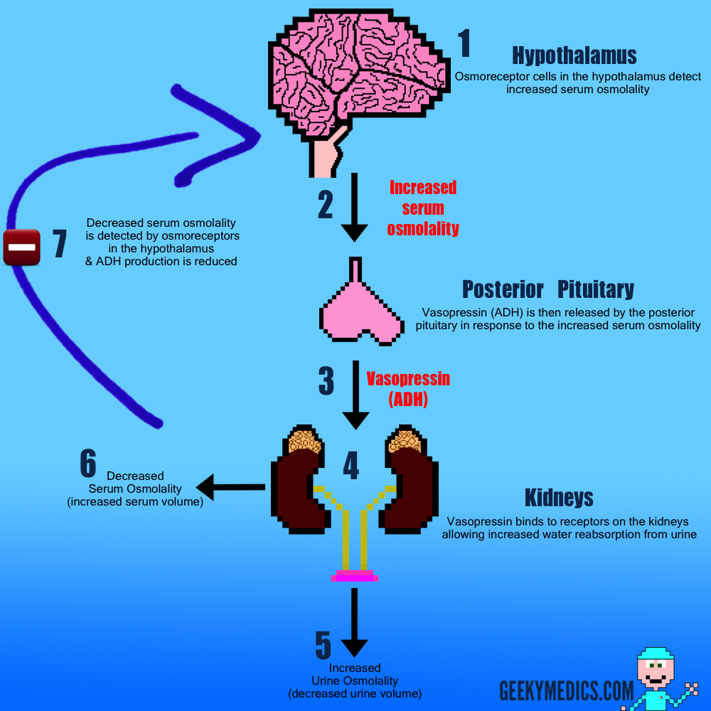
1. Vasopressin (Anti-Diuretic Hormone) is produced by the hypothalamus in response to increased serum osmolality

2. Vasopressin is then transported to the posterior pituitary gland

3. It is then released into the circulatory system via the posterior pituitary gland

4. It then travels to the kidneys where it binds to vasopressin receptors on the distal convoluted tubules

5. This causes Aquaporin-2 channels to move from the cytoplasm into the apical membrane of the tubules.

  • These aquaporin-2 channels allow water to be reabsorbed out of the collecting ducts & back into the blood stream.
  • This results in both a decrease in volume & an increase in osmolality (concentration) of the urine been excreted

6. The extra water that has been reabsorbed re-enters the circulatory system, reducing the serum osmolality

7. This reduction in serum osmolality is detected by the hypothalamus & results in decreased production of vasopressin.

Causes
Neurogenic

Diabetes insipidus can occur as a result of decreased circulating levels of Vasopressin (ADH). Vasopressin is responsible for instructing the kidneys to retain fluid.  Therefore decreased circulating levels of ADH results in the production of copious volumes of urine.  Because vasopressin is produced by the hypothalamus & released by the posterior pituitary gland, pathology impacting either of these glands has the potential to cause diabetes insipidus.

.

Familial

Mutations in the Vasopressin gene   (e.g. Autosomal dominant AVP-NPII)¹

  • Results in inadequate production of functional Vasopressin

.

Acquired¹

Tumours – Pituitary adenoma’s (20%), Craniopharyngiomas, Metastases
Trauma – 17% of cases - e.g. Head injury
Neurosurgery – 9% of cases
Infections – meningitis
Vascular – Sheehan’s syndrome
  • Complication of pregnancy in which the pituitary blood supply is ↓ causing necrosis of the gland
Sarcoidosis – formation of granulomas in pituitary
Haemochromotosis - deposition of iron in pituitary/hypothalamic tissue causing damage
Langerhans’ cell histiocytosis -
  • Proliferation of Langerhans cells which form lesions in many organs including pituitary stalk³
Idiopathic – 25% of cases.
Nephrogenic

The kidneys are responsible for reabsorbing fluid when ADH binds to their receptors.  Anything which interferes with this binding or damages the kidneys has the potential to cause diabetes insipidus.


Familial

X-linked recessive – mutations in the ADH receptor gene¹

Autosomal recessive – aquaporin-2 gene – aquaporin 2 is responsible for the reabsorption of water from urine4

..

Acquired 

Metabolic – hypercalcaemia, hyperglycaemia, hypokalaemia
Drugs – lithium, demeclocycline – both interfere with the binding of ADH¹
Chronic renal disease – polycystic kidney’s
Amyloidosis
Post obstructive uropathy¹

.

Dipsogenic

Dipsogenic diabetes insipidus is caused by a defect or damage to the hypothalamus causing malfunction of the thirst mechanism¹.  As a result the individual is excessively thirsty regardless of their fluid status.  The individual therefore consumes large volumes of fluid which suppresses secretion of vasopressin and increases urine output.  It is dangerous to give a vasopressin analogue such as Desmopressin in these circumstances as the individual will continue to feel thirsty and consume large volumes of fluids which could result in fluid overload.


Gestational

Gestational diabetes insipidus only occurs during pregnancy.  During pregnancy the placenta produces vasopressinase which breaks down vasopressin.  Gestational diabetes insipidus is therefore thought to be caused by overproduction of vasopressinase by the placenta causing a lack of functional vasopressin.


Primary Polydipsia 

Primary polydipsia is characterised by an individual consuming large volumes of fluids and as a result producing large volumes of dilute urine.  The symptoms of primary polydipsia are therefore very similar to those of diabetes insipidus however a fluid deprivation test can help distinguish the diseases.  Most often primary polydipsia is due to a psychological disorder.


Symptoms & Signs
Symptoms

Excessive urination (>3L/24hrs)

Excessive thirst (especially for ice cold water)

Nocturia

Dehydration – headache, dizziness, fainting, dry mouth

Signs

Hypotension

Dilute urine

Reduced capillary refill time

Investigations

Measure urine output – confirm more than 3000ml a day

Exclude diabetes mellitus – dipstick urine for glucose & assess blood glucose level

Exclude renal failure

.

Check electrolyte levels

  • Hypokalaemia & Hypercalcaemia – nephrogenic DI¹
  • Hypernatraemia can develop due to dehydration

.

Fluid deprivation test¹
  • Helps determine cause of DI – neurogenic, nephrogenic, primary polydipsia
  • Patient is allowed fluids overnight
  • Patient is then deprived of fluids for 8 hours (or until loss of 5% of body weight if earlier)
  • The patient is weighed hourly
  • Plasma osmolality is measured every 4 hours
  • Urine volume & Osmolality is measured every 2 hours
  • At the end of the deprivation period the patient is given 2mcg of IM Desmopressin
  • Urine volume & Serum osmolality are then measured over the next 4 hours

.

MRI – scan head looking for tumours - pituitary adenomas, craniopharyngoma’s
Diagnosis

The fluid deprivation test is the most useful in diagnosing diabetes insipidus

It can confirm the presence of DI and suggest which type of DI the individual likely has

If the serum osmolality is >305mOsm/kg at any point the patient has DI (stop test)


Table demonstrating patterns of urine osmolality in the fluid deprivation test and their corresponding likely diagnosis¹

Neurogenic

If the diagnosis is Neurogenic DI the urine osmolality will be low after fluid deprivation but normalise after desmopressin is given. This is because neurogenic DI is caused by the lack of vasopressin production therefore giving a synthetic form of vasopressin such as desmopressin normalises levels of the hormone resulting in the normalisation of serum & urine osmolality.

.

Nephrogenic

If the diagnosis is Nephrogenic DI then the urine osmolality will remain low throughout regardless of desmopressin.  This is because the kidneys have a problem which prevents them from been able to respond to vasopressin, therefore giving extra synthetic vasopressin will have no effect.

.

Primary Polydipsia

If the diagnosis is Primary Polydipsia the urine osmolality will remain high after fluid deprivation as well as after desmopressin is given.  This is because the patient’s vasopressin axis is intact and otherwise completely normal.

.

Partial DI or Polydipsia

If the diagnosis is that of Partial DI or Polydipsia the picture may be mixed.  The patient may have a slightly low osmolality after fluid deprivation and may not reach normal urine osmolality after desmopressin.  This kind of picture would require more thorough investigation to determine a definitive cause.


Management
Neurogenic

Give Desmopressin¹

  • Vasopressin analogue
  • Binds to v2 receptors on kidney allowing water to be reabsorbed
  • Drug can be given orally, intranasally, parenterally or bucally
  • Dose varies significantly between patients
  • Osmolality & Serum Sodium need monitoring – can cause hyponatraemia or hypo-osmolality

Chlorpropamide & Carbamazepine - can be used to increase activity of vasopressin¹


Nephrogenic

Advise patient to maintain adequate fluid intake

Correct any metabolic derangement’s  hypercalcaemia, hyperglycaemia, hypokalaemia¹

Stop any drugs that may be to blame -  lithium, demeclocycline - both interfere with the binding of ADH¹

High dose Desmopressin – up to 5mcg IM

Thiazide diuretics / Prostaglandin Synthase Inhibitors – reduce action of prostaglandins which can inhibit  vasopressin’s action on the kidney¹


Partial Diabetes Insipidus

Advise patient to maintain adequate fluid intake - usually no drugs required

  • Thirst mechanism must be normal¹
  • Polyuria must be mild (<4L/24hrs)

.

Primary Polydipsia

Often very difficult to manage

The underlying psychiatric disorder needs to be treated 


References

Click to show

1. Oxford handbook of Endocrinology & Diabetes

2. Saborio, P.; Tipton, G. A.; Chan, J. C. M. (2000). “Diabetes Insipidus”. Pediatrics in Review 21 (4): 122–129

3. Makras P, Papadogias D, Kontogeorgos G, Piaditis G, Kaltsas G (2005). “Spontaneous gonadotrophin deficiency recovery in an adult patient with Langerhans cell histiocytosis (LCH)”. Pituitary 8 (2): 169–74

4. Wildin, Robert (2006). What is NDI?. The Diabetes Inspidus Foundation


Syndrome of inappropriate antidiuretic hormone secretion

$
0
0
What is SIADH?

The syndrome of inappropriate antidiuretic hormone secretion (SIADH) involves the excessive secretion of antidiuretic hormone (ADH) from the posterior pituitary gland or another source. ADH controls water reabsorption by the kidneys nephrons, causing the retention of water but not solute. Therefore ADH causes dilution of the blood which decreases the concentrations of solutes such as sodium.

Physiology
Normal

1. Vasopressin (Anti-Diuretic Hormone) is produced by the hypothalamus in response to increased serum osmolality

2. Vasopressin is then transported to the posterior pituitary gland

3. It is then released into the circulatory system via the posterior pituitary gland

4. It then travels to the kidneys where it binds to vasopressin receptors on the distal convoluted tubules

5. This causes Aquaporin-2 channels to move from the cytoplasm into the apical membrane of the tubules.

  • These aquaporin-2 channels allow water to be reabsorbed out of the collecting ducts & back into the blood stream.
  • This results in both a decrease in volume & an increase in osmolality (concentration) of the urine been excreted

6. The extra water that has been reabsorbed re-enters the circulatory system, reducing the serum osmolality

7. This reduction in serum osmolality is detected by the hypothalamus & results in decreased production of vasopressin.

ADH Metabolism

Deranged physiology in SIADH

SIADH has a number of potential causes. These are demonstrated in the diagram below. The important difference in SIADH, is the lack of any negative feedback mechanism, resulting in an inability to reduce or stop ADH production. As a result, ADH is continually produced, regardless of what the serum osmolality is. This ultimately results in abnormally low levels of serum sodium and relatively high levels of urinary sodium,  giving rise to the characteristic symptoms and signs associated with SIADH.

SIADH Derranged physiology.

What causes SIADH?

Brain damage – Meningitis, Sub-arachnoid haemorrhage (SAH)

MalignancySmall-cell lung cancer

Drugs – Carbamazepine, SSRI’s, Amitriptyline, Morphine

Infective – Pneumonia, Lung abscess, Brain abscess

Hypothyroidism

Signs & Symptoms
Symptoms¹

Symptoms vary greatly depending on the timescale of the development of hyponatraemia

Therefore mild hyponatraemia may cause significant symptoms if the drop in sodium is acute whereas chronically hyponatraemic patients may have very low serum sodium concentrations and yet be completely asymptomatic. This is thought to be due to cerebral adaptation, where brain cells can adapt their metabolism to cope with abnormal sodium levels, but this can only occur if the change in sodium concentration is gradual.


Mild – Nausea / Vomiting / Headache / Anorexia / Lethargy

Moderate – Muscle cramps / Weakness / Confusion / Ataxia

Severe – Drowsiness / Seizures / Coma

Signs¹

These also vary a great deal depending on the rate of serum sodium concentration change

  • Decreased GCS
  • Cognitive impairment (short term memory loss, disorientation, confusion)
  • Focal or generalised seizures
  • Brain stem herniation - severe acute hyponatraemia - coma, respiratory arrest
  • Hypervolaemia -pulmonary oedema, peripheral oedema, raised JVP, Ascites


Investigations³

Fluid status:

- Is the patient clinically or biochemically dehydrated?
- In SIADH this is not the case – the patient is either euvolemic or hypervolaemic
- If dehydration is present, it suggests another cause for hyponatraemia – diuretics, renal failure etc


Serum Sodium - low in SIADH -  <135 mmol/L

Serum Potassium - if raised in the presence of hyponatraemia consider Addison’s

Plasma Osmolality – this will also be reduced due to the low sodium concentration


Urine Osmolality:

- Normally if serum osmolality is low, urine osmolality should also be

- This is because the kidneys should be trying to retain solute

- In SIADH the excess ADH prevents the kidney from retaining solute

- As a result concentrated urine, high in Na, is produced, despite low serum Na


Urine Sodium 

- This will be relatively raised, in the context of the serum sodium concentration
- This is because the excess ADH prevents the kidney from retaining sodium


TFT’s - hypothyroidism is a cause of SIADH – ↓ T3 & ↑TSH would suggest this diagnosis

Serum Cortisol - Addison’s causes ↓ Na, a low serum cortisol would suggest this diagnosis

Imaging - useful in detecting causes of SIADH such as small cell lung Ca, SAH e.g. CT head


Algorithm to determine the cause of hyponatraemia

Algorithm to determine the cause of hyponatraemia²

Diagnosis

The following features need to be present for a diagnosis of SIADH:³

  • Hyponatraemia
  • LowPlasma Osmolality
  • Inappropriately elevated urine osmolality (>plasma osmolality)
  • Urine [Na+] >40 mmol/L with normal salt intake
  • Euvolaemia
  • Normal Thyroid & Adrenal Function
Management

It’s difficult to give a generic step by step management strategy for SIADH, as it can be due to so many different causes. As a result, you need to target your management strategy at the underlying cause for long term correction of sodium metabolism.


Fluid restriction

This is a common management strategy for increasing serum sodium concentrations, at least temporarily, whilst the underlying cause is sought and treated. Usually the fluid restriction is between 1-1.5 litres per day. It’s largely dependent on patients co-operating with the treatment plan, which some patients can struggle with.


Replacing Sodium

Another general management strategy for treating hyponatraemia is to replace sodium by giving normal saline 0.9% as a slow infusion. This has to be done with great care, as if the sodium concentration is corrected too rapidly it can result in the devastating complication known as Central Pontine Myelinolysis. This is characterised by permanent damage to the myeline sheath in the brain stem, causing acute paralysis, dysphagia, dysarthria, diplopia and loss of consciousness. As a result, this treatment strategy has to be done with extreme caution, with the recommendation of not correcting serum sodium levels more than 10 mmol/L/24h.


Demeclocycline 

This drug is not first line and only used when fluid restriction alone fails to resolve hyponatraemia. Demeclocycline is a tetracycline antibiotic that reduces the sensitivity of the ADH receptors on the distal tubules of the kidneys. As a result this partially blocks the effects of ADH on the kidneys, essentially creating a partial nephrogenic diabetes insipidus.


Vaptans

Vaptans are  a new class or drugs known as vasopressin receptor antagonists

Tolvaptan is a V2 selective vasopressin receptor antagonist that is sometimes used to treat resistant hyponatraemia in SIADH, Heart Failure & Liver Cirrhosis

It selectively blocks the effects of ADH (vasopressin) on the kidneys

This drug is for specialist use only, given it’s price & the potential for side effects such as liver injury & excessive thirst.


Examples of treating the underlying cause

Pneumoniaantibiotics

Carbamezipineconsider switch to alternative anti-epileptic i.e. Sodium Valproate

Hypothyroidism - levothyroxine replacement

Small cell lung cancer – chemotherapy

References

Click to show

1. Craig S; Hyponatremia in Emergency Medicine, Medscape, Apr 2010 (Symptoms & Signs)

2. Oxford handbook of clinical medicine 7th edition [p666-667]

3. Robert D. Zenenberg,Do, et. al (2010-04-27). ”Hyponatremia: Evaluation and Management”. Hospital Practice. 38 (1): 89–96


Hyperparathyroidism

$
0
0
What is Hyperparathyroidism?

Hyperparathyroidism is characterised by excessive production of parathyroid hormone (PTH) by the parathyroid glands. There are 4 of these glands in the neck, located behind the thyroid gland. The glands themselves are small, been around the size of your little finger nail.  PTH is normally secreted in response to low ionised calcium levels. PTH promotes increased Osteoclast activity, causing the release of Calcium and Phosphate into the serum. It also acts on the kidneys to increase calcium and decrease phosphate reabsorption at the proximal tubule. The end result of producing too much PTH is therefore hypercalcaemia, hypophosphataemia and loss of bone density.¹

Classification of Hyperparathyroidism
Primary

Primary Hyperparathyroidism is due to excessive production of PTH by one or more of the parathyroid glands. This is usually a result of a small PTH secreting adenoma within the gland.


Secondary

Secondary Hyperparathyroidism is the result of a normal physiological response to hypocalcaemia, which may be due to a myriad of different causes such as:

  • Chronic renal failure (commonest cause)
  • Chronic Pancreatitis
  • Small bowel malabsorption


Tertiary

Secretion of PTH autonomously - often as a result of chronic kidney disease

Physiology

1. Low serum calcium is detected by the parathyroid glands

2. This promotes production of parathyroid hormone (PTH)

3. PTH is released into the blood

4. PTH promotes osteoclast activity in the bones

5. Osteoclasts dissolve bone & the calcium from this moves into the serum

6. PTH also ↑ calcium reabsorption by the kidneys & ↓ phosphate reabsorption

7. PTH also promotes the conversion of Vitamin D in the kidney into it’s more active form

8. ↑ levels of active Vitamin D promote increased reabsorption of calcium in the bowel

9. Bone resorption alongside ↑ renal & bowel reabsorption results in ↑ serum calcium levels

10. Increased serum calcium levels inhibit further production of PTH via negative feedback

PTH Axis


Investigations
Bloods

CalciumThis is raised in all forms of hyperparathyroidism

Parathyroid Hormone - PTH is also raised in all types of hyperparathyroidism

Phosphatethis is low in all types of  hyperparathyroidism due to the effect of PTH on the kidneys

U&E’smay show ↑creatinine suggesting chronic renal disease – a common cause of 2° & tertiary hyperparathyroidism


Review patients medications for drugs promoting hypercalcaemia
  • Lithium
  • Thiazide diuretics


Imaging

DEXA scan

A DEXA scan measures bone mineral density (BMD)

Patients with hyperparathyroidism often have low BMD due to the effect of PTH on osteoclasts

A DEXA is therefore useful in assessing the severity of the disease as well as response to treatment

It’s results are invaluable when considering management strategies


MIBI scan 

This scan is used in suspected primary hyperparathyroidism

The aim is to identify PTH secreting adenomas in the parathyroid glands

The contrast used is a complex of  technetium-99m with the ligand methoxyisobutylisonitrile (MIBI)

The parathyroid gland takes up this complex after intravenous injection

The patient’s neck is then imaged with a gamma camera to show the location of all glands.

A second image is obtained after a washout time (approximately 2 hours)

Abnormal PTH secreting adenoma glands retaining the 99mTc and are seen with the gamma camera

The test can detect 75% to 90% of abnormal parathyroid glands in primary hyperparathyroidism

Localisation of the abnormal gland allows surgeons to do a less invasive directed parathyroidectomy


Neck USS

An USS scan of the parathyroid glands is often done in addition to the MIBI scan

It looks for enlarged parathyroid glands suggestive of adenoma

Use of both USS & MIBI can increase the sensitivity & specificity of identifying single adenomas

The biggest issue is that Neck USS is far more operator dependent than a MIBI scan

So the USS operator skill significantly influences the usefulness of the test


Renal tract imaging

Patients who are hypercalcaemic are significantly more likely to develop renal stones

This can often be the way hyperparathyroidism first presents

As a result, those who are diagnosed with hyperparathyroidism often have a KUB x-ray performed

This will demonstrate any potential stones

Recurrent renal stones can lead to irreversible renal damage & pre-dispose to potential urosepsis


Biopsy

Parathyroid biopsies are sometimes performed if carcinoma is suspected

Carcinoma is rare however.


Primary Hyperparathyroidism

Primary Hyperparathyroidism is due to ↑ production of PTH by 1 or more of the parathyroid glands.

This is usually a result of a small PTH secreting adenoma within the gland.

It is the 3rd most common endocrinological disease (incidence of 1 in a 1000)

It is 3 times more common in women than men

Post-menopausal women have the highest incidence


Symptoms / Signs

Most symptoms & signs occur as a result of the hypercalcaemia

The pneumonic “Stones, Bones, Abdominal Groans & Psychiatric Moans” is a useful aide-mémoire


Stones 

Kidney stonesstones made of calcium salts which can block the urinary tract
Nephrocalcinosisdeposition of calcium in the renal parenchyma – can lead to renal failure
Acquired Diabetes Insipidus hypercalcaemia causes increased sodium loss in the urine & therefore loss of more water with it


Bones

Osteoporosis:

  • Excessive PTH promotes the destruction of bone by osteoclasts
  • As a result bone mineral density decreases


Osteitis fibrosa cystica:

  • Bone pain - especially the long bones e.g. Tibia 
  • Peritrabecular fibrosis - replacement of bone with fibrous tissue
  • Brown cyst-like tumours form throughout the skeleton


Abdominal Groans

Constipation 
Indigestion
Nausea & Vomiting
Peptic ulcers
Pancreatitis


Psychiatric Moans

Lethargy
Fatigue
Depression
Poor memory
Delirium
Psychosis


Diagnosis

Serum Calcium - ↑

Parathyroid Hormone - ↑

U&E’srenal function may be normal or impaired if hypercalcaemia has been longstanding

MIBImay show a single enlarged parathyroid gland suggestive of adenoma

DEXAdepends on length & severity of disease – usually less than normal


Management
Conservative

This approach is often used in mild/asymptomatic disease

This is defined as elevated Ca & relatively normal bone / renal investigations

A conservative approach involves:

  • Check Calcium levels every 6 months
  • Also check Creatinine levels to identify renal damage early
  • Annual DEXA scan
  • Advise high fluid intake – keeps calcium lower & reduces risk of renal stones
  • Moderate calcium intake


Surgical

If patient has any of the following, the surgery is usually advised:

  • Symptomatic hypercalcaemia
  • Significant loss of bone density
  • Renal stones


Single parathyroid gland demonstrated to be hyperplastic or contain adenoma on MIBI scan:

  • Directed Parathyroidectomy – Targeted removal of the causative glands 
  • This is a less invasive procedure – only a small incision needed
  • This leads to improved cosmetic results


Single gland can not be identified / or all glands are thought to be hyperplastic:

  • More invasive procedure involving an incision over the thyroid is performed
  • The scar is similar to that of a thyroidectomy
  • In this procedure three and a half of the four glands are removed
  • The remaining half of a parathyroid gland will provide sufficient levels of PTH to control calcium levels adequately (ideally)


Complications of surgery


Hypocalcaemia

This is due to the phenomena often referred to as hungry bone syndrome.  Because of the rapid fall in PTH levels after surgery, it’s inhibitory effect on osteoblasts is suddenly reversed.  As a result osteoblasts rapidly cause absorption of calcium from the serum to produce new bone. This results in a serum drop in Calcium.  After a while the remaining normal parathyroid glands adjust their sensitivity & produce more PTH which corrects this issue.


Haematoma formation

This is a risk of any surgery, however due to the anatomical location of the glands there is significant risk associated with haematomas.  A haematoma in the pre-tracheal space can rapidly result in airway occlusion.  If this is suspected post-operatively, emergency management involves immediate removal of sutures.  This allows pressure to be relieved. Further surgery to stop bleeding may be required.


Recurrent laryngeal nerve injury

Another significant risk of the surgery.

The nerve runs very close to the operative site, therefore damage can occur

This results in a hoarse voice.

Often this resolves a few weeks post-op, but in some cases it can persist permanently


Medical

Medical treatment is not first line in the majority of cases

It is reserved for those whom surgery is not appropriate

Or alternatively for those who refuse to have surgery

A combination of medications is used with the aim to improve / maintain bone mineral density whilst decreasing calcium to normal levels


Cinacalcet

Cinacalcet is a calcimimetic (mimics action of calcium, by binding to the same receptor)
It therefore binds to the calcium sensing receptors on the parathyroid glands
This results in a negative feedback effect reducing the secretion of PTH
This causes reduction of calcium levels & an increase in phosphate levels
It is recommended for the management of secondary hyperparathyroidism


Bisphosphonates

Bisphosphonates such as Alendronate are another drug used in Hyperparathyroidism.
They protect from bone loss & can lead to an increase in bone density.
They work by promoting apoptosis of osteoclasts, which are responsible for breaking down bone.
As a result bone loss is reduced.


Secondary Hyperparathyroidism
Cause

Secondary hyperparathyroidism occurs as a result of chronic hypocalcaemia

Because of continous stimulation, the parathyroid glands become hyperplastic

The most common causes of chronic hypocalcaemia are:

  • Renal failure
  • Malabsorption
  • Vitamin D deficiency
Diagnosis

Hypocalcaemia

Raised PTH

Phosphate - raised in renal disease – low in vitamin D deficiency

Management

Medical management is the first line approach in secondary hyperparathyroidism

Treatment of the underlying pathology causing hypocalcaemia is the main focus


Vitamin D deficiency:

  • Replacement of Vitamin D with supplements


Chronic Kidney Disease:

  • Caclium & Vitamin D supplements (e.g. Calcichew D3)
  • Cinacalcet – only recommended in end stage renal disease in which other treatment has failed


Tertiary Hyperparathyroidism
Causes

Most commonly tertiary hyperparathyroidism develops after prolonged secondary hyperparathyroidism. The parathyroid glands eventually stop paying attention to blood calcium levels & therefore the negative feedback mechanism of reducing PTH production is lost. As a result, even when serum calcium levels are restored to normal, the glands continue to produce excessive volumes of PTH, resulting in hypercalcaemia. The most common cause of this is chronic kidney disease.

Investigation

Hypercalcaemia

Raised PTH

Raised Phosphate

Management

Parathyroidectomy is the 1st line management -  total / subtotal parathyroidectomy

This is because the parathyroid glands have become  autonomous

So altering PTH production via medical methods is usually ineffective

References

Click to show

1. Fraser WD; Hyperparathyroidism. Lancet. 2009 Jul 11;374(9684):145-58.

Hyperthyroidism

$
0
0
What is Hyperthyroidism?

Hyperthyroidism is a disease process caused by the presence of an overactive thyroid gland which in turn results in excessive production of thyroid hormones (T3 & T4)

When hyperthyroidism becomes symptomatic it is referred to as thyrotoxicosis

Random Fact Thyroid comes from the Greek “threoeides” which means shield shaped

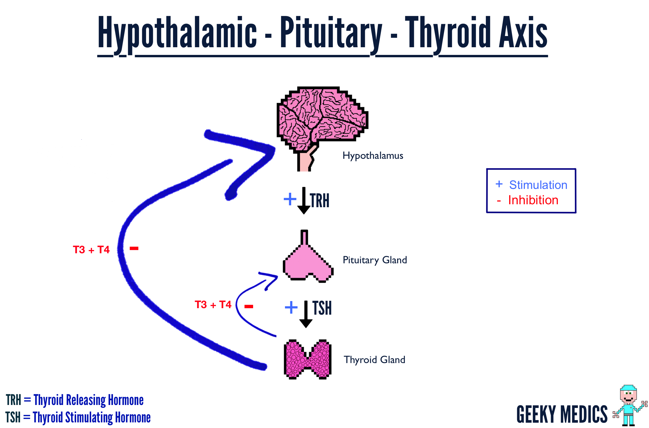
Hypothalamic-Pituitary-Thyroid Axis


.

Causes
Graves Disease

Graves’ disease is the most common cause of hyperthyroidism.  It is an autoimmune disease directed against Thyroid Stimulating Hormone (TSH) receptors.  The autoantibodies stimulate the TSH receptors causing ↑ T3 & T4 production.  It is most common in middle aged women, with a female to male ratio of 8:1.  Patients become symptomatic due to thyrotoxicosis (high levels of T3 & t4).  It can be diagnosed by the presence of thyrotoxicosis and thyroid-stimulating hormone receptor (TSH-R) antibodies which are specific to Graves’ disease.


Toxic Multinodular Goitre

Caused by hyperplasia of groups of thyroid cells.  The increased number of cells can result in increased production of thyroid hormones .


Toxic Thyroid Adenoma

A benign tumour of the thyroid gland which produces excessive amounts of thyroid hormones.  These always arise from the follicular cells of the thyroid.  Not all thyroid adenomas are toxic (capable of producing T3 & T4) these are often referred too as “cold” thyroid adenomas.


Thyroiditis

Inflammation of the thyroid gland due to infections or lymphocytic infiltration causes the stored thyroid hormones to all be released into the circulation leading to hyperthyroidism and usually thyrotoxicosis. However because of the inflammation the thyroid is unable to produce thyroid hormone effectively, this results in the patient developing hypothyroidism in the long termHashimoto’s thyroiditis, an autoimmune disease against thyroglobulin (T3 & T4 precursor), is an example of this process.


Drug Induced

Particular drugs can cause an increase in the levels of thyroid hormones via many mechanisms.

Thyroxine overdose is probably the most common.

Amiodarone is capable of causing hyperthyroidism as well as hypothyroidism.

 
Hamburger Hyperthroidism

This form of hyperthyroidism is caused by consumption of ground beef which has been contaminated with thyroid tissue.  The thyroid tissue contains metabolically active thyroid hormones which cause the hyperthyroidism.  This is something you wont likely ever need to know, however it’s the kind of thing consultants love to ask.

Thyroid Storm (hyperthyroid crisis)

Severe hyperthyroidism causing:
  • Fever
  • Tachycardia
  • Atrial Fibrillation
  • Diarrhoea & Vomiting
  • Agitation
  • Confusion
  • Coma

It can be caused by recent thyroid surgery, radio-iodine, infection, trauma

Aggressive treatment is required as the condition is life threatening – seek specialist help

Symptoms & Signs
Symptoms

Heat intolerance

Weight loss

Palpitations

Hair loss

Anxiety / Restlessness

Diarrhoea

Increased appetite

Menorrhagia

Reduced libido

Signs

Goitre

Thyroid Nodules

Proximal Myopathy

Hyperreflexia

Fine Hand Tremor

Pre-tibial myxoedema

Tachycardia

Atrial Fibrillation


Eyes

– Exopthalmos (protrusion of eyes)
– Lid retraction
– Lid lag

Investigations
Thyroid function tests

Main method of diagnosis:

  • Serum TSH
  • Free T3
  • Free T4
 
A high T3 & T4 confirm presence of hyperthyroidism
If TSH is low the problem is likely with the thyroid gland itself
If TSH is high it suggests a problem in the pituitary gland or higher up the chain
Autoantibodies

Thyroglobulin antibody (Tg Ab) – present in Graves & Hashimoto’s
Thyroid Peroxidase antibody (TPO Ab) – present in Graves & Hashimoto’s
Thyroid-stimulating hormone receptor antibodies (TSH-R) – specific to Graves 

ECG

Check for presence of atrial fibrillation


Isotope Scan

Visualises thyroid

Allows estimation of size

Allows areas of increased activity to be seen

Treatment
Carbimazole

Blocks synthesis of T3 & T4

Therefore lowering active circulating thyroid hormones

 
Radioiodine

Thyroid is only gland to take up iodine

Radiation causes thyroid cell necrosis

A significant number of patients develop hypothyroidism & need thyroxine for life

The patient cannot be in close contact with others until 2 weeks after treatment due to radiation

 
Thyroidectomy

Removal of thyroid tissue to reduce production of T3 & T4

Risk of damaging recurrent laryngeal nerve (causing hoarse voice)

Risk of hypoparathyroidism (due to parathyroid glands close relation to thyroid tissue)

 
Beta Blocker

Control atrial fibrillation

Therefore reducing risk of emboli formation

Prognosis

Treatment is successful in most patients

Most require yearly monitoring of thyroid hormones

Some patients may relapse into a hyperthyroid state years later

The post Hyperthyroidism appeared first on Geeky Medics.

Hypothyroidism

$
0
0
What is Hypothyroidism?

Hypothyroidism refers to the presence of an under-active thyroid gland which causes a deficiency of thyroid hormones (T3 & T4).  When hypothyroidism becomes symptomatic it is referred to as myxoedema. It’s prevalence is slightly lower than that of hyperthyroidism, affecting around 1% of females and 0.2% of males. The disease often has an insidious onset with unspecific symptoms, so it’s important to be remain aware, especially in those older than 40 and female.

Hypothalamic-Pituitary-Thyroid Axis


.
Causes
Hashimoto’s Thyroiditis

Hashimoto’s thyroiditis is the most common cause of hypothyroidism

It is a destructive autoimmune disease that is especially common in middle aged women.

The disease is caused by auto-reactive antibodies against  thyroglobulin & other targets

The process begins with lymphocytic infiltration of the thyroid, giving the characteristic goitre

This can cause the release of stored thyroid hormones causing a transient episode of hyperthyroidism

Eventually the inflammation & destruction of the thyroid gland result in hypothyroidism

 
De Quervain’s Thyroiditis

De Quervain’s Thyroiditis is inflammation of the thyroid gland as the result of a virus

The disease is often preceded by a upper respiratory tract infection

The most common pathogens involved include coxsackie virus, mumps and adenoviruses

It most often affects young & middle aged women

It tends to cause a tender swollen thyroid gland in combination with fever

 
Primary Atrophic Hypothyroidism

Primary atrophic hypothyroidism generally only affects the elderly.

It occurs as the result of  atrophy & fibrosis of the gland.

This condition can also be the end result of diseases such as Hashimoto’s Thyroiditis.

 
Iodine Deficiency

Iodine is an essential component to thyroid hormones and therefore lack of it causes hypothyroidism.

Iodine deficiency also result in the formation of a goitre

This is due to excessive TSH been produced by the pituitary in response to low T3 & T4.

The high levels of TSH causes the thyroid glands tissues to grow to soak up any available iodine.

This results in a characteristic smooth diffusely enlarged goitre.

Iodine deficiency is now very rare in developed nations due to iodination of salt.

Iodine deficiency in developing children can cause severe mental & growth retardation

.

Random FactA town called Derby in England had very low levels of iodine in the soil and as a result there was a very high prevalence of goitre which was given the name “Derbyshire Neck”


Iatrogenic Hypothyroidism

Often people treated for hyperthyroidism subsequently develop hypothyroidism

This occurs due to destruction or inhibition of the thyroid tissue

Patients are therefore regularly monitored during treatment

 
Postpartum Thyroiditis

Postpartum thyroiditis is inflammation of the thyroid following pregnancy

It often causes initial hyperthyroidism which often may return to normal

However some patients subsequently develop hypothyroidism

1 in 5 of these women will have the disease lifelong

The disease is believed to develop due to changes in the immune system necessary for pregnancy

This results in inappropriate immune activation against thyroid tissue

 
Congenital Hypothyroidism

Congenital hypothyroidism involves the deficiency of the thyroid hormones at birth

It occurs in approximately 1 in 4000 children

All children in the UK are screened at birth

It can be due to a number of reasons including; prematurity, genetic conditions & pituitary function

The treatment is to replace the thyroid hormones using thyroxine which is cheap and effective

Children treated early will develop normally and live an otherwise normal life

Symptoms & Signs

The disease often goes unnoticed at first due to it’s insidious onset

Symptoms

Fatigue

Decreased appetitive

Weight gain

Cold Intolerance

Dry skin

Hair Loss

Constipation

Menorrhagia / Amenorrhea

 
Signs

Decreased tendon reflexes

Cramps / Spasms

Carpal Tunnel Syndrome

Blurry Vision

Deepening of voice

Pale skin

Peripheral Neuropathy

Peri-orbital oedema

Pericardial effusion

Depression

Psychosis

Encephalopathy

Coma

Investigations
Thyroid function tests;

Main diagnosis is based on:

  • Free T3 & T4
  • Serum TSH

A low T3 & T4 confirm presence of hypothyroidism

If TSH is high the problem is likely with the thyroid gland itself

If TSH is low it suggests a problem in the pituitary gland or higher up the chain


Antithyroid Antibodies

Antibody testing can be useful in determining the cause of hypothyroidism

Thyroglobulin antibody (Tg Ab) present in Graves & Hashimoto’s
Thyroid Peroxidase antibody (TPO Ab)present in Graves & Hashimoto’s

Antithyroid microsomal antibody – antibodies against rough ER

Thyroid Stimulating Hormone Receptor Antibody (TSH Ab)

Treatment
Thyroxine

Almost all forms of hypothyroidism can be treated very effectively with Thyroxine

It comes in tablet form and is relatively cheap and available

Overdose can result in thyrotoxicosis

Prognosis

Prognosis is excellent with thyroxine therapy (as long as patient complies with treatment regime)

The post Hypothyroidism appeared first on Geeky Medics.

Acromegaly

$
0
0
What is Acromegaly?

Acromegaly is a disease caused by the overproduction of growth hormone (GH) after an individual has reached adulthood and epiphyseal plate (growth plate) closure has occurred.  This overproduction of GH is as a result of a pituitary tumour in more than 99% of cases, however in very rare circumstances it can caused by ectopic production of GH by carcinoid tumours.  Acromegaly is a rare condition affecting approximately 60 in a million people.  It’s prevalence is distributed equally between males & females.  It tends to affect those between 30-50 years old.

Mechanism

The disease process usually starts with the development of a benign pituitary adenoma.
The adenoma begins to produce excessive amounts of growth hormone (GH)
↑ GH stimulates increased production of Insulin-like Growth Factor 1 (IGF1) by the liver
IGF1 is the hormone responsible for stimulating growth of tissues
As a result organs, bone, muscle & other tissues begin to overgrow
Increased levels of IGF1 also cause changes in blood glucose control & lipid metabolism
The ↑IGF1 also often causes hypertension
If the disease remains untreated overgrowth of myocardial tissue results in ventricular hypertrophy and eventually heart failure

Symptoms & Signs
Symptoms

Headache
Excessive sweating
Hoarse voice
Sleep Apnea – laryngeal soft tissue growth
Arthralgia
Carpal Tunnel Syndrome
Visual changes – due to compression of optic chiasm by pituitary adenoma

Signs

Enlargement of hands – ↑ ring size, broad spade like hands

Enlargement of feet – ↑shoe size

Changes in facial features;

– Thickened lips & nose
– Increased jaw prominence (prognathism)
– Prominent supraorbital ridge
– Wide spaced teeth
– Large tongue (macroglossia)
– Thickened oily skin


Visual field defects – Bi-temporal heminopia

Proximal Muscle Weakness

Tinel’s Sign Positive – carpal tunnel syndrome

Diagnosis
Serum IGF-1

Serum IGF-1 is the test used to screen for Acromegaly

The levels of IGF-1 directly relate to the amount of GH secretion over the previous 24hrs

Therefore if IGF-1 is found to be raised it suggests large amounts of GH secretion

Unfortunately in 25% of cases IGF-1 is normal even thought GH is raised leading to false negatives

 
Oral Glucose Tolerance Test (OGTT)

The gold standard test is an Oral Glucose Tolerance Test (OGTT) combined with GH measurement

GH secretion is normally inhibited by ↑ glucose & should therefore be undetectable

In Acromegaly the production of GH is not affected by glucose & therefore will be raised

 
Other Tests

MRI of Pituitary Fossa – performed if previous tests suggest presence of Acromegaly

Visual Fields – pituitary tumours can often compress the close by optic chiasm

Visual Acuity

ECG – cardiac hypertrophy?

Echocardiogram – cardiac hypertrophy?

Older photos of patient – changes in facial features?

Treatment
Trans-sphenoidal Surgery

Direct removal of pituitary adenoma

Cure rate of:

– 80% in micro-adenoma
– 40% in macro-adenoma 

GH & Pituitary function monitored afterwards

If GH remains high other therapies may be needed

 
Somatostatin Analogues

Inhibit release of GH

Examples Octreotide & Lanreotide

Administered every 2-4 weeksinjection

Most patients respond well

GH levels often falls within an hour of treatment & headaches can resolve in minutes

Can cause GI disturbance & loose stoolsmay not be well tolerated

 
Growth Hormone Receptor Antagonists

This drug type is a new development & is very targeted

They block the action of the endogenous growth hormone molecules

The drug is called Pegvisomant

It’s administered subcutaneously by daily injections

The exciting thing is this treatment is able to control disease activity in virtually all patients


Radiotherapy

Used if surgery is inappropriate

Can be used as an adjuvant if tumour remaining after surgery

Can take a long time for effects to be seen (years in some cases)

The post Acromegaly appeared first on Geeky Medics.

How the adrenal axis works

$
0
0

What is the adrenal axis?

The adrenal axis (hypothalamic-pituitary-adrenal axis) refers to a complex set of interactions and feedback loops between the hypothalamus, pituitary and adrenal glands. This system regulates the body’s response to stress, immune function, energy expenditure, mood, emotions and libido. 

What are the adrenal glands?

  • There is an adrenal gland located above each kidney
  • They produce a number of different hormones
  • The adrenal glands are composed of an outer cortex and an inner medulla

Adrenal cortex

The adrenal cortex is responsible for producing cortisol and aldosterone.

Cortisol 

  • Cortisol is a steroid hormone (glucocorticoid)
  • It is released in response to stress and low blood-glucose concentration.
  • Some of its functions include:
    • Increasing blood glucose levels by promoting gluconeogenesis
    • Suppressing the immune system
    • Aiding the metabolism of fat, protein and carbohydrates


Aldosterone

  • Aldosterone is also a steroid hormone (mineralocorticoid)
  • Its main function is to increase blood volume
  • It causes reabsorption of sodium and water, in addition to excretion of potassium

Adrenal medulla

  • The adrenal medulla produces adrenaline and noradrenaline (both have similar actions).
  • They are released as an acute response to stress and imitate many of the effects of cortisol
  • They essentially prepare the body for “fight or flight”↑HR,  ↑BP,  ↑blood flow to muscles
  • Release is under direct control from the hypothalamus
  • Adrenaline and noradrenaline have a much shorter half life than cortisol

How cortisol is regulated

  1. Corticotropic releasing hormone (CRH) is secreted from the hypothalamus
  2. This release is influenced by stress levels, time of day and serum cortisol levels
  3. CRH travels in the blood and binds to specific receptors in the pituitary gland
  4. This binding causes increased production of adrenocorticotropic hormone (ACTH)
  5. ACTH is released into the bloodstream where it travels to the adrenal glands
  6. ACTH binds to specific receptors in the adrenal cortex
  7. This stimulates the adrenal cortex to release cortisol into the blood
  8. Cortisol enables the body to cope with stress in a more effective manner
  9. Increased levels of cortisol also have an immunosuppressive effect
  10. Blood glucose is also increased via breakdown of glycogen, protein and fat
  11. Increased serum cortisol inhibits production of CRH and ACTH via negative feedback


.
.

How aldosterone is regulated

1. Low blood volume triggers release of angiotensin II.
2. Angiotensin II stimulates the adrenal cortex to produce aldosterone.

Aldosterone has a number of different actions:

  • Sodium reabsorption
  • Water reabsorption
  • Potassium excretion

3. Aldosterone’s actions result in a net increase in blood volume.
4. Increased blood volume inhibits production of Angiotensin II via negative feedback.


The post How the adrenal axis works appeared first on Geeky Medics.

How the gonadal axis works

$
0
0

What is the gonadal axis?

The gonadal axis involves a complex interaction between the hypothalamus, pituitary gland and the gonads.  The system works together to regulate development, reproductionageing and many other body processes.  Its regulation relies upon a number of complex negative feedback loops which when lost, result in disease.

How the male gonadal axis works

1. The hypothalamus secretes GnRH.

2. GnRH travels down to the anterior pituitary gland.

3. It binds to receptors on the pituitary gland.

4. This causes release of LH (luteinizing hormone) and FSH (follicle stimulating hormone).

5. LH and FSH travel in the blood stream to the testicles.


6. LH stimulates leydig cells in the testicles to produce testosterone
  • Testosterone is required for spermatogenesis and many other important biological processes


7. FSH stimulates sertoli cells to produce androgen binding globulin (ABG) and inhibin
  • ABG is a protein which binds to testosterone and keeps it within the seminiferous tubules
  • Inhibin helps support spermatogenesis and inhibits production of FSH, LH and GnRH

8. levels of testosterone and inhibin cause negative feedback on the pituitary and hypothalamus.

9. This results in decreased production of LH and FSH.

10. As a result production of testosterone and inhibin is also decreased.


How the female gonadal axis works

1. The hypothalamus secretes GnRH.

2. GnRH travels down to the anterior pituitary gland.

3. It binds to receptors on the pituitary gland.

4. This causes release of LH (luteinizing hormone) and FSH (follicle stimulating hormone).

5. LH and FSH travel in the blood stream to the ovaries.

6. When LH and FSH bind to the ovaries they stimulate production of oestrogen and inhibin

  • Oestrogen helps regulate the menstrual cycle and is essential in many body processes
  • Inhibin causes inhibition of activin which is usually responsible for stimulating GnRH production

7. Increasing levels of oestrogen and inhibin cause negative feedback on the pituitary and hypothalamus.

8. This leads to decreased production of GnRH, LH and FSH.

9. This in turn results in decreased production of oestrogen and inhibin.


The post How the gonadal axis works appeared first on Geeky Medics.


The Menstrual Cycle

$
0
0

What is the menstrual cycle?

The menstrual cycle is a complex series of physiological changes occurring in women on a monthly basis.  It results in production of an ovum and thickening of the endometrium to allow for implantation if fertilisation should occur.  The menstrual cycle is orchestrated by the endocrine system through the complex interaction of the hypothalamus, pituitary and gonads.  The entire cycle lasts around 28 days, with the cycle beginning on the first day of menstruation and ovulation occurring around day 14.

How is it controlled?

1. The hypothalamus produces Gonadotrophin Releasing Hormone (GnRH).

2. This binds to the pituitary stimulating release of:

  • Luteinizing hormone (LH)
  • Follicle Stimulating Hormone (FSH)

.

3. FSH binds to the ovaries stimulating:

  • Development of  ovarian follicles
  • Secretion of oestrogen
  • Secretion of inhibin

The follicle most sensitive to FSH becomes dominant and is known as the Graafian follicle

.

4. LH binds to the ovaries causing:

  • Production of oestrogen which is required for ovulation and thickening of the endometrium
  • Conversion of the Graafian follicle into the progesterone producing corpus luteum
  • Progesterone causes the endometrium to become receptive to implantation of a fertilised ovum

.

5. Oestrogen, Progesterone and Inhibin all cause negative feedback on the pituitary and hypothalamus.

6. This results in reduction of GnRH, FSH and LH production.

7. In pregnancy GnRH, FSH and LH all remain inhibited, causing cessation of menstruation.

Phases of the menstrual cycle

Follicular phase

1. At the start of the cycle levels of FSH rise causing stimulation of a few ovarian follicles.

2. As follicles mature they compete with each other for dominance.

3. The 1st follicle to become fully mature will produce large amounts of oestrogen.

4. This inhibits the growth of the other competing follicles.

5. The 1 follicle reaching full maturity is called the Graafian follicle (oocyte develops within this).

6. The Graafian follicle continues to secrete increasing amounts of oestrogen.

.

7. Oestrogen causes:

  • Endometrial thickening
  • Thinning of cervical mucous to allow easier passage of sperm

.

8. Oestrogen also initially inhibits LH production from the pituitary gland.

9. However when the ovum is mature, oestrogen reaches a threshold level which conversely causes a sudden spike in LH around day 12.

10. The high amounts of LH cause the membrane of the Graafian follicle to become thinner.

11. Within 24-48 hours of the LH surge, the follicle ruptures releasing a secondary oocyte.

12. The secondary oocyte quickly matures into an ootid and then into a mature ovum.

13. The ovum is then released into the peritoneal space and is taken into the fallopian tube via fimbriae (finger like projections).

Luteal phase

14. Once ovulation has occurred the hormones LH and FSH cause the remaining graafian follicle to develop into the corpus luteum.
15. The corpus luteum then begins to produce the hormone progesterone.

.

16. Increased levels of progesterone result in:

  • Endometrium becoming receptive to implantation of the blastocyst
  • Increased production of oestrogen by the adrenal glands
  • Negative feedback causing decreased LH and FSH (both needed to maintain the corpus luteum)
  • Increase in the woman’s basal body temperature
    .

17. As the levels of FSH and LH fall, the corpus luteum degenerates.

18. This results in progesterone no longer being produced.

18. The falling level of progesterone triggers menstruation and the entire cycle starts again.

19. However if an ovum is fertilised it produces hCG which is similar in function to LH.

20. This prevents degeneration of the corpus luteum (continued production of progesterone).

21. Continued production of progesterone prevents menstruation.

22. The placenta eventually takes over the role of the corpus luteum (from 8 weeks).

The Uterine Cycle

The uterus has its own cycle which is driven by the cyclical release of hormones by the ovaries which we’ve previously talked about.  The inside lining of the uterus is known as the endometrium. The endometrium is the part of the uterus most affected by these changes in hormone levels.

.

It is composed of 2 layers:

  • Functional layer – this grows thicker in response to oestrogen and is shed during menstruation
  • Basal layer – this forms the foundation from which the functional layer develops – it is not shed

Phases of the uterine cycle

The uterine cycle has 3 phases known as the proliferative, secretory and menstrual phases.

Proliferative phase

During the proliferative phase the endometrium is exposed to an increase in oestrogen levels caused by FSH and LH stimulating the ovaries. This oestrogen causes repair and growth of the functional endometrial layer allowing recovery from the recent menstruation and further proliferation of the endometrium.

.

Continued exposure to increasing levels of oestrogen causes:

  • Increased endometrial thickness
  • Increased vascularity –spiral arteries grow into the functional endometrial layer
  • Development of increased numbers of secretory glands

.

Secretory phase

The secretory phase begins once ovulation has occurred.

This phase is driven by progesterone produced by the corpus luteum.

It results in the endometrial glands beginning to secrete various substances.

These secretions make the uterus a more welcoming environment for an embryo to implant.

.

Menstrual phase

At the end of the luteal phase the corpus luteum degenerates (if no implantation occurs).

The loss of the corpus luteum results in decreased progesterone production.

The decreasing levels of progesterone cause the spiral arteries in the functional endometrium to contract.

The loss of blood supply causes the functional endometrium to become ischaemic and necrotic.

As a result the functional endometrium is shed and exits out through the vagina.

This is seen as the 3-5 day period of menstruation a woman experiences each month.

Window of fertility

A woman’s most fertile period is between 5 days before ovulation until 1 to 2 days after.

Women can therefore use knowledge of their cycle to improve chances of conception.

Symptoms experienced in the menstrual cycle

Abdominal pain and cramps

Vaginal bleeding

Nausea

Diarrhoea

Sweating

Fatigue

Irritability

Dysphoria (unhappiness)

The post The Menstrual Cycle appeared first on Geeky Medics.

Diabetes insipidus

$
0
0

What is diabetes insipidus?

Diabetes insipidus is a disease characterised by the passage of large volumes (>3L/24hrs) of dilute urine (osmolality <300 mOsmol/Kg)¹. It affects approximately 3 in 100,000 people². In some cases the volume of urine produced can be as much as 20 litres in a 24 hour period and therefore rapid dehydration can easily occur, leading to death if not managed appropriately. Diabetes insipidus has a number of causes and therefore use of the correct investigations is essential to reach a definitive diagnosis.

Pathophysiology

1. Vasopressin (antidiuretic hormone) is produced by the hypothalamus in response to increased serum osmolality.

2. Vasopressin is then transported to the posterior pituitary gland.

3. Vasopressin is then released into the circulatory system via the posterior pituitary gland.

4. Vasopressin then travels to the kidneys where it binds to vasopressin receptors on the distal convoluted tubules.

5. This causes aquaporin-2 channels to move from the cytoplasm into the apical membrane of the tubules:

  • These aquaporin-2 channels allow water to be reabsorbed out of the collecting ducts and back into the bloodstream.
  • This results in both a decrease in volume and an increase in osmolality (concentration) of the urine being excreted.

6. The extra water that has been reabsorbed re-enters the circulatory system, reducing the serum osmolality.

7. This reduction in serum osmolality is detected by the hypothalamus and results in decreased production of vasopressin.

Causes

Neurogenic

Diabetes insipidus can occur as a result of decreased circulating levels of vasopressin (ADH). Vasopressin is responsible for instructing the kidneys to retain fluid, therefore decreased circulating levels of ADH results in the production of copious volumes of urine.  Vasopressin is produced by the hypothalamus and released by the posterior pituitary gland, meaning damage to either of these structures can cause diabetes insipidus.

.

Familial

Mutations in the Vasopressin gene   (e.g. Autosomal dominant AVP-NPII)¹

  • Results in inadequate production of functional Vasopressin

.

Acquired¹

Tumours – Pituitary adenomas (20%) / Craniopharyngiomas / Metastases
Trauma – 17% of cases
Neurosurgery – 9% of cases
Infections – meningitis
Vascular – Sheehan’s syndrome
  • Complication of pregnancy in which the pituitary blood supply is ↓ causing necrosis of the gland
Sarcoidosis – formation of granulomas in pituitary
Haemochromotosis – deposition of iron in pituitary/hypothalamic tissue causing damage
Langerhans’ cell histiocytosis – proliferation of Langerhans cells which form lesions in many organs including pituitary stalk³
Idiopathic – 25% of cases.

Nephrogenic

The kidneys are responsible for reabsorbing fluid when antidu binds to their receptors.  Anything which interferes with this binding or damages the kidneys has the potential to cause diabetes insipidus.


Familial

X-linked recessive – mutations in the ADH receptor gene¹

Autosomal recessive – aquaporin-2 gene – aquaporin 2 is responsible for the reabsorption of water from urine 4

..

Acquired 

Metabolic – hypercalcaemia / hyperglycaemia / hypokalaemia
Drugs – lithium / demeclocycline – both interfere with the binding of ADH¹
Chronic renal disease
Amyloidosis
Post obstructive uropathy¹

Dipsogenic

Dipsogenic diabetes insipidus occurs as a result of hypothalamic disease or trauma. The hypothalamus is responsible for controlling thirst which is a key component of maintaining an appropriate fluid balance¹. As a result the individual is excessively thirsty regardless of their fluid status.  The individual therefore consumes large volumes of fluid which suppresses secretion of vasopressin and increases urine output.  It is dangerous to give a vasopressin analogue such as desmopressin in these circumstances as the individual will continue to feel thirsty and consume large volumes of fluids which could result in fluid overload.


Gestational

Gestational diabetes insipidus only occurs during pregnancy.  During pregnancy the placenta produces vasopressinase which breaks down vasopressin.  Gestational diabetes insipidus is therefore thought to be caused by overproduction of vasopressinase by the placenta causing a lack of functional vasopressin.


Primary polydipsia 

Primary polydipsia is characterised by an individual consuming large volumes of fluids and as a result producing large volumes of dilute urine.  The symptoms of primary polydipsia are therefore very similar to those of diabetes insipidus however a fluid deprivation test can help distinguish the diseases.  Most often primary polydipsia is due to a behavioural disorder.


Symptoms and signs

Symptoms

Excessive urination (>3L/24hrs)

Excessive thirst (especially for ice cold water)

Nocturia

Dehydration – headache / dizziness / dry mouth

Signs

Hypotension

Dilute urine

Reduced capillary refill time

Investigations

Measure urine output – confirm more than 3000ml a day

Exclude diabetes mellitus – dipstick urine for glucose and assess blood glucose level

Exclude renal failure – check U&Es

.

Check electrolyte levels

  • Hypokalaemia and Hypercalcaemia – nephrogenic DI¹
  • Hypernatraemia can develop due to dehydration

.

Fluid deprivation test¹
  • Helps determine cause of DI – neurogenic / nephrogenic / primary polydipsia
  • Patient is allowed fluids overnight
  • Patient is then deprived of fluids for 8 hours (or until loss of 5% of body weight if earlier)
  • The patient is weighed hourly
  • Plasma osmolality is measured every 4 hours
  • Urine volume and osmolality is measured every 2 hours
  • At the end of the deprivation period the patient is given 2mcg of intramuscular desmopressin
  • Urine volume and serum osmolality are then measured over the next 4 hours

.

MRI head – pituitary adenomas / craniopharyngiomas

Diagnosis

  • The fluid deprivation test is the most useful in diagnosing diabetes insipidus (DI)
  • It can confirm the presence of DI and suggest which type of DI the individual likely has
  • If the serum osmolality is >305mOsm/kg at any point the patient has DI (stop test)


Table demonstrating patterns of urine osmolality in the fluid deprivation test and their corresponding likely diagnosis¹

Neurogenic

If the diagnosis is Neurogenic DI the urine osmolality will be low after fluid deprivation but normalise after desmopressin is given. This is because neurogenic DI is caused by the lack of vasopressin production therefore giving a synthetic form of vasopressin such as desmopressin normalises levels of the hormone resulting in the normalisation of serum and urine osmolality.

.

Nephrogenic

If the diagnosis is nephrogenic DI then the urine osmolality will remain low throughout regardless of desmopressin.  This is because the kidneys have a problem which prevents them from being able to respond to vasopressin.

.

Primary polydipsia

If the diagnosis is primary polydipsia the urine osmolality will remain high after fluid deprivation as well as after desmopressin is given.  This is because the patient’s vasopressin axis is intact and otherwise completely normal.

.

Partial DI or polydipsia

If the diagnosis is that of partial DI or polydipsia the picture may be mixed.  The patient may have a slightly low osmolality after fluid deprivation and may not reach normal urine osmolality after desmopressin.  This kind of picture would require more thorough investigation to determine a definitive cause.


Management

Neurogenic

Desmopressin¹

  • Vasopressin analogue
  • Binds to v2 receptors on kidney allowing water to be reabsorbed
  • Drug can be given orally, intranasally, parenterally or buccally
  • Dose varies significantly between patients
  • Serum osmolality and serum sodium need monitoring – can cause hyponatraemia or hypo-osmolality


Nephrogenic

Advise patient to maintain adequate fluid intake

Correct any metabolic derangements –  hypercalcaemia, hyperglycaemia, hypokalaemia¹

Stop any drugs that may be to blame –  lithium, demeclocycline – both interfere with the binding of ADH¹

High dose desmopressin

Thiazide diuretics / prostaglandin synthase inhibitors – reduce action of prostaglandins which can inhibit the action of vasopressin on the kidneys¹.


Partial diabetes insipidus

Advise patient to maintain adequate fluid intake – usually no drugs required

  • Thirst mechanism must be normal¹
  • Polyuria must be mild (<4L/24hrs)

.

Primary polydipsia

Often very difficult to manage.

The underlying psychiatric disorder needs to be treated .

References

Click to show

1. Oxford handbook of Endocrinology & Diabetes

2. Saborio, P.; Tipton, G. A.; Chan, J. C. M. (2000). “Diabetes Insipidus”. Pediatrics in Review 21 (4): 122–129

3. Makras P, Papadogias D, Kontogeorgos G, Piaditis G, Kaltsas G (2005). “Spontaneous gonadotrophin deficiency recovery in an adult patient with Langerhans cell histiocytosis (LCH)”. Pituitary 8 (2): 169–74

4. Wildin, Robert (2006). What is NDI?. The Diabetes Inspidus Foundation

The post Diabetes insipidus appeared first on Geeky Medics.

Syndrome of inappropriate antidiuretic hormone secretion (SIADH)

$
0
0

What is SIADH?

The syndrome of inappropriate antidiuretic hormone secretion (SIADH) involves the excessive secretion of antidiuretic hormone (ADH) from the posterior pituitary gland or another source. ADH controls water reabsorption by the kidneys nephrons, causing the retention of water but not solute. Therefore ADH causes dilution of the blood which decreases the concentration of solutes such as sodium.

Physiology

Normal

1. Vasopressin (Anti-Diuretic Hormone) is produced by the hypothalamus in response to increased serum osmolality.

2. Vasopressin is then transported to the posterior pituitary gland.

3. It is then released into the circulatory system via the posterior pituitary gland.

4. It then travels to the kidneys where it binds to vasopressin receptors on the distal convoluted tubules.

5. This causes Aquaporin-2 channels to move from the cytoplasm into the apical membrane of the tubules:

  • These aquaporin-2 channels allow water to be reabsorbed out of the collecting ducts and back into the bloodstream.
  • This results in both a decrease in volume and an increase in osmolality (concentration) of the urine being excreted.

6. The extra water that has been reabsorbed re-enters the circulatory system, reducing the serum osmolality.

7. This reduction in serum osmolality is detected by the hypothalamus and results in decreased production of vasopressin.

ADH Metabolism

Deranged physiology in SIADH

SIADH has a number of potential causes. These are demonstrated in the diagram below. The important difference in SIADH, is the lack of any negative feedback mechanism, resulting in an inability to reduce or stop ADH production. As a result, ADH is continually produced, regardless of what the serum osmolality is. This ultimately results in abnormally low levels of serum sodium and relatively high levels of urinary sodium, giving rise to the characteristic symptoms and signs associated with SIADH.


.

What causes SIADH?

Brain damage – Meningitis / Subarachnoid haemorrhage (SAH)

MalignancySmall-cell lung cancer

Drugs – Carbamazepine / SSRIs / Amitriptyline

Infectious – Atypical pneumonia / Lung abscess /Cerebral abscess

Hypothyroidism

Signs and Symptoms

Symptoms¹

Symptoms vary greatly depending on the timescale of the development of hyponatraemia

Therefore mild hyponatraemia may cause significant symptoms if the drop in sodium is acute whereas chronically hyponatraemic patients may have very low serum sodium concentrations and yet be completely asymptomatic. This is thought to be due to cerebral adaptation, where brain cells can adapt their metabolism to cope with abnormal sodium levels, but this can only occur if the change in sodium concentration is gradual.


Mild – Nausea / Vomiting / Headache / Anorexia / Lethargy

Moderate – Muscle cramps / Weakness / Confusion / Ataxia

Severe – Drowsiness / Seizures / Coma

Signs¹

These also vary a great deal depending on the rate of serum sodium concentration change

  • Decreased level of consciousness 
  • Cognitive impairment (short term memory loss / disorientation / confusion)
  • Focal or generalised seizures
  • Brain stem herniation severe acute hyponatraemia (coma / respiratory arrest)
  • Hypervolaemiapulmonary oedema / peripheral oedema / raised JVP / ascites


Investigations³

Fluid status:

  • Is the patient clinically or biochemically dehydrated?
  • In SIADH this is not the case – the patient is either euvolemic or hypervolaemic
  • If dehydration is present, it suggests another cause for hyponatraemia – diuretics / renal failure

Serum sodium – low in SIADH –  <135 mmol/L

Serum potassium – if raised in the presence of hyponatraemia consider Addison’s

Plasma osmolality – this will also be reduced due to the low sodium concentration


Urine osmolality:

  • Normally if serum osmolality is low, urine osmolality should also be low
  • This is because the kidneys should be trying to retain solute
  • In SIADH the excess ADH causes water retention but not solute retention
  • As a result concentrated urine relatively high in sodium is produced, despite low serum sodium


Urine sodium :

  • This will be relatively raised, in the context of the serum sodium concentration


TFTs – hypothyroidism is a cause of SIADH – ↓ T3 & ↑TSH would suggest this diagnosis

Serum cortisol – Addison’s causes ↓ Na, a low serum cortisol would suggest this diagnosis

Imaging – useful in detecting causes of SIADH such as small cell lung Ca


Algorithm to determine the cause of hyponatraemia

Algorithm to determine the cause of hyponatraemia²

Diagnosis

The following features need to be present for a diagnosis of SIADH:³

  • Hyponatraemia
  • Low plasma osmolality
  • Inappropriately elevated urine osmolality (>plasma osmolality)
  • Urine [Na+] >40 mmol/L with normal salt intake
  • Euvolaemia
  • Normal thyroid and adrenal function

Management

It’s difficult to give a generic step by step management strategy for SIADH, as it can be due to so many different causes. As a result, you need to target your management strategy at the underlying cause for long term correction of sodium metabolism. Specialist endocrine input should be sought before commencing any of these management strategies.


Fluid restriction

This is a common management strategy for increasing serum sodium concentrations, at least temporarily, whilst the underlying cause is sought and treated. Usually the fluid restriction is between 1-1.5 litres per day. It’s largely dependent on patients co-operating with the treatment plan, which some patients can struggle with.


Replacing sodium

Another general management strategy for treating hyponatraemia is to replace sodium either orally or via IV fluid. This has to be done with great care, as if the sodium concentration is corrected too rapidly it can result in the devastating complication known as central pontine myelinolysis. This is characterised by permanent damage to the myelin sheath in the brain stem, causing acute paralysis, dysphagia, dysarthria, diplopia and decreased level of consciousness. As a result, this treatment strategy has to be carried out with extreme caution, with the recommendation of not correcting serum sodium levels more than 10 mmol/L/24h.


Examples of treating the underlying cause

Pneumoniaantibiotics

Carbamazepineconsider switch to alternative anti-epileptic i.e. sodium valproate

Hypothyroidism levothyroxine replacement

References

Click to show

1. Craig S; Hyponatremia in Emergency Medicine, Medscape, Apr 2010 (Symptoms & Signs)

2. Oxford handbook of clinical medicine 7th edition [p666-667]

3. Robert D. Zenenberg,Do, et. al (2010-04-27). “Hyponatremia: Evaluation and Management”. Hospital Practice. 38 (1): 89–96

The post Syndrome of inappropriate antidiuretic hormone secretion (SIADH) appeared first on Geeky Medics.

How the adrenal axis works

$
0
0

What is the adrenal axis?

The adrenal axis (hypothalamic-pituitary-adrenal axis) refers to a complex set of interactions and feedback loops between the hypothalamus, pituitary and adrenal glands. This system regulates the body’s response to stress, immune function, energy expenditure, mood, emotions and libido. 


What are the adrenal glands?

  • There is an adrenal gland located above each kidney
  • They produce a number of different hormones
  • The adrenal glands are composed of an outer cortex and an inner medulla

Adrenal cortex

The adrenal cortex is responsible for producing cortisol and aldosterone.

Cortisol 

  • Cortisol is a steroid hormone (glucocorticoid)
  • It is released in response to stress and low blood-glucose concentration.
  • Some of its functions include:
    • Increasing blood glucose levels by promoting gluconeogenesis
    • Suppressing the immune system
    • Aiding the metabolism of fat, protein and carbohydrates


Aldosterone

  • Aldosterone is also a steroid hormone (mineralocorticoid)
  • Its main function is to increase blood volume
  • It causes reabsorption of sodium and water, in addition to excretion of potassium

Adrenal medulla

  • The adrenal medulla produces adrenaline and noradrenaline (both have similar actions).
  • They are released as an acute response to stress and imitate many of the effects of cortisol
  • They essentially prepare the body for “fight or flight”↑HR,  ↑BP,  ↑blood flow to muscles
  • Release is under direct control from the hypothalamus
  • Adrenaline and noradrenaline have a much shorter half life than cortisol

How cortisol is regulated

  1. Corticotropic releasing hormone (CRH) is secreted from the hypothalamus
  2. This release is influenced by stress levels, time of day and serum cortisol levels
  3. CRH travels in the blood and binds to specific receptors in the pituitary gland
  4. This binding causes increased production of adrenocorticotropic hormone (ACTH)
  5. ACTH is released into the bloodstream where it travels to the adrenal glands
  6. ACTH binds to specific receptors in the adrenal cortex
  7. This stimulates the adrenal cortex to release cortisol into the blood
  8. Cortisol enables the body to cope with stress in a more effective manner
  9. Increased levels of cortisol also have an immunosuppressive effect
  10. Blood glucose is also increased via breakdown of glycogen, protein and fat
  11. Increased serum cortisol inhibits production of CRH and ACTH via negative feedback


.
.

How aldosterone is regulated

1. Low blood volume triggers release of angiotensin II.
2. Angiotensin II stimulates the adrenal cortex to produce aldosterone.

Aldosterone has a number of different actions:

  • Sodium reabsorption
  • Water reabsorption
  • Potassium excretion

3. Aldosterone’s actions result in a net increase in blood volume.
4. Increased blood volume inhibits production of Angiotensin II via negative feedback.


The post How the adrenal axis works appeared first on Geeky Medics.

How the gonadal axis works

$
0
0

What is the gonadal axis?

The gonadal axis involves a complex interaction between the hypothalamus, pituitary gland and the gonads.  The system works together to regulate development, reproductionageing and many other body processes.  Its regulation relies upon a number of complex negative feedback loops which when lost, result in disease.


How the male gonadal axis works

1. The hypothalamus secretes GnRH.

2. GnRH travels down to the anterior pituitary gland.

3. It binds to receptors on the pituitary gland.

4. This causes release of LH (luteinizing hormone) and FSH (follicle stimulating hormone).

5. LH and FSH travel in the blood stream to the testicles.


6. LH stimulates leydig cells in the testicles to produce testosterone
  • Testosterone is required for spermatogenesis and many other important biological processes


7. FSH stimulates sertoli cells to produce androgen binding globulin (ABG) and inhibin
  • ABG is a protein which binds to testosterone and keeps it within the seminiferous tubules
  • Inhibin helps support spermatogenesis and inhibits production of FSH, LH and GnRH

8. levels of testosterone and inhibin cause negative feedback on the pituitary and hypothalamus.

9. This results in decreased production of LH and FSH.

10. As a result production of testosterone and inhibin is also decreased.


How the female gonadal axis works

1. The hypothalamus secretes GnRH.

2. GnRH travels down to the anterior pituitary gland.

3. It binds to receptors on the pituitary gland.

4. This causes release of LH (luteinizing hormone) and FSH (follicle stimulating hormone).

5. LH and FSH travel in the blood stream to the ovaries.

6. When LH and FSH bind to the ovaries they stimulate production of oestrogen and inhibin

  • Oestrogen helps regulate the menstrual cycle and is essential in many body processes
  • Inhibin causes inhibition of activin which is usually responsible for stimulating GnRH production

7. Increasing levels of oestrogen and inhibin cause negative feedback on the pituitary and hypothalamus.

8. This leads to decreased production of GnRH, LH and FSH.

9. This in turn results in decreased production of oestrogen and inhibin.


The post How the gonadal axis works appeared first on Geeky Medics.

Viewing all 51 articles
Browse latest View live